 |
"If I only had a
brain...."
A primer on the brain and cranial anatomy of mosasaurs
(With apologies to a certain Scarecrow from the Land of Oz)
Copyright © 2008-2010 by Mike Everhart
Page created 09/08/2008; Last updated 01/01/2010
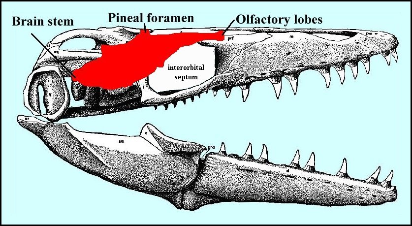
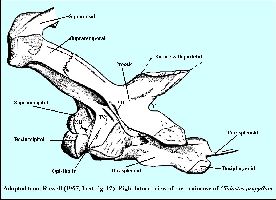
LEFT: The skull of Platecarpus
ictericus in right lateral view, with the location of the brain in red. Adapted from
Russell (1967, Text-fig. 37), with kudos to Mike Polcyn for his knowledgable
assistance. |
Mosasaurs obviously had a brain...maybe not a very big one by mammalian
standards, but certainly large enough to allow them to rule as top predators in the
Cretaceous seas for millions of years.
The most difficult part of the mosasaur skull for me to understand (and
visualize) is the back one-third, including the braincase. With rare exceptions, mosasaur
skulls are crushed fairly flat when collected and many of the structural relationships
between the individual bones are lost or not readily apparent. While this not a major
problem with understanding the tooth bearing structures in front, the three dimensional
aspect of the back of the skull is difficult to reconstruct. Note that in many reptiles,
including mosasaurs, the braincase is cartilagenous and does not preserve as a fossil
(with possible rare exceptions). In addition, there is a thin sheet of cartilage, called
the interorbital septum, that separates the eyeballs. The pineal foramen is the mosasaur
brain's window to the outside world... often called a "third eye" and is
associated with the pineal gland. The olfactory lobes run between and above the eyes
and nest under the frontal bone where they are in contact with the nasal passages.
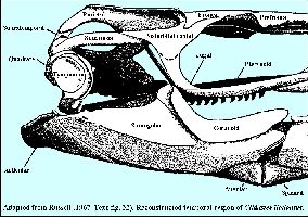
I've adapted a series of figures from the mosasaur bible (Russell, 1967) that
help you understand which bones make up the braincase or are connected it. The large open
spaces between the bones, especially those areas on either side of the parietal and behind
the squamosals were filled with strong muscles that attached to the lower and provided the
means for quickly and powerfully closing the jaws.
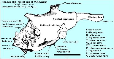 |
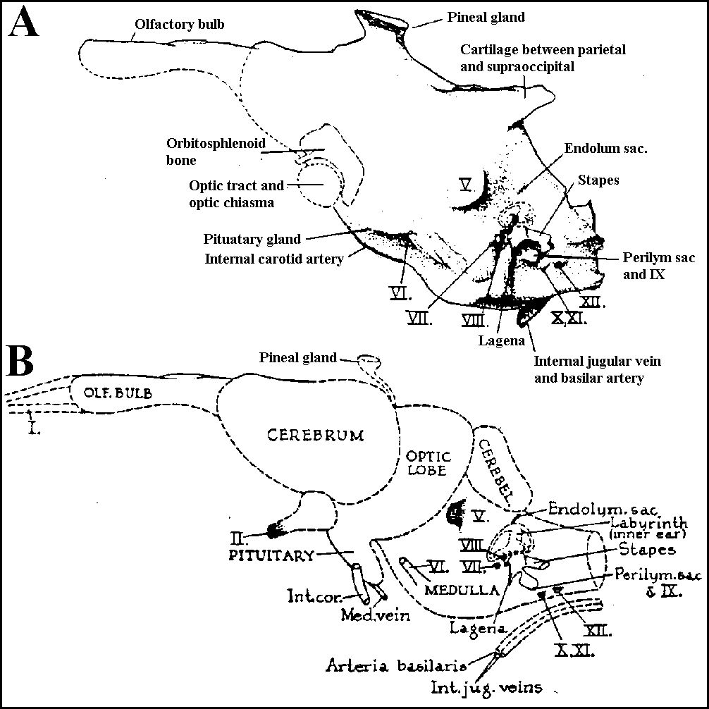
LEFT: Starting with the general shape of the brain
(from an endocast).... and location of the cranial nerves - the spinal cord (and
vertebrae) would be to the left. The olfactory lobe (sense of smell) was located along the
underside of the frontal, between the large eyeballs It was relatively small in
size, not unusual for marine animals. The eyes were quite large, probably useful for
seeing in dimly lit or deeper waters, and indicate that mosasaurs probably were sight
hunters. However, since the eyes were located on the side of the head, they did not have
stereoscopic vision. Like most animals, including humans, the mosasaur brain has
twelve pairs of cranial nerves. Cranial nerves III and IV (not shown) control the
movements of the eyes. Cranial nerve VIII is responsible for hearing and balance. |
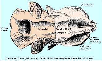 |
LEFT: The floor of the mosasaur braincase is composed
of two main bones: the basioccipital, which connects to the atlas/axis vertebrae, and the
basisplenoid. Most of the blood supply for the brain (basilar and internal carotid
arteries) comes through these two bones. These and a cartilageenous structure enclose the
brain in something more like a tunnel than the rounded cranium that most of us are
familiar with in our own skull. |
 |
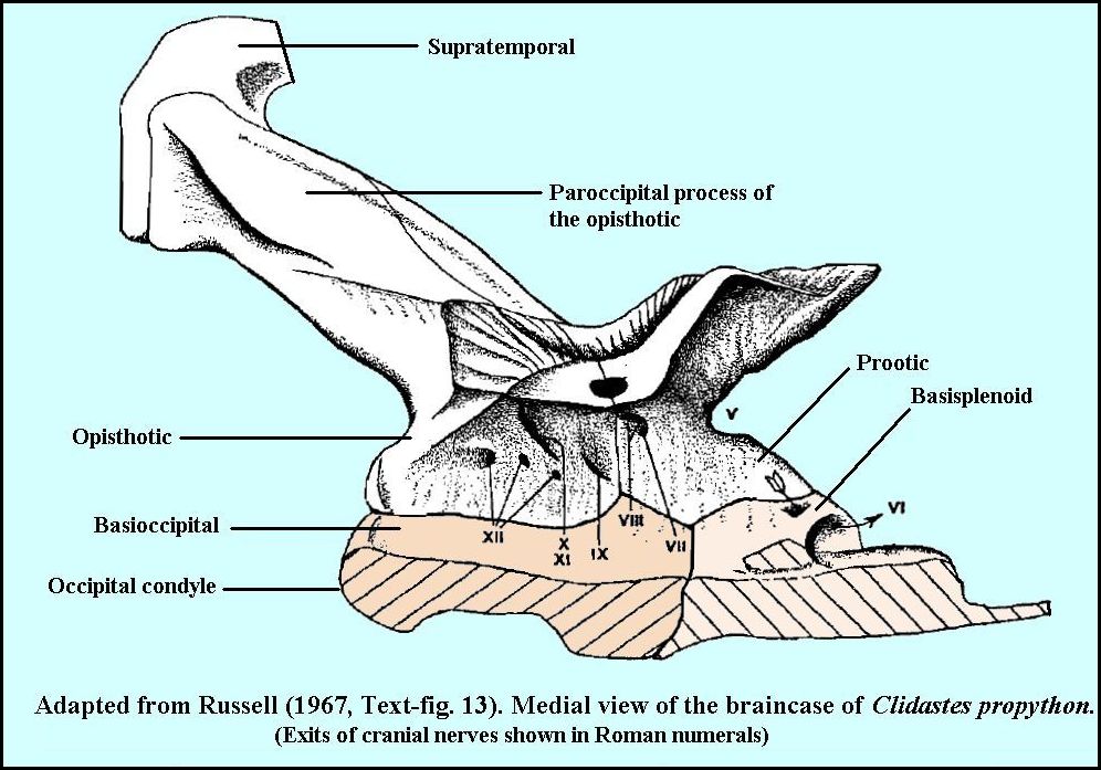
LEFT: Next we have a medial view of the braincase, showing an interior
view of the bones that enclose the mosasaur brain. Basioccipital and basisplenoid below,
paired opisthotic and prootic above. These bones are penetrated by or enclose openings
(foramina) for the cranial nerves. |
 |
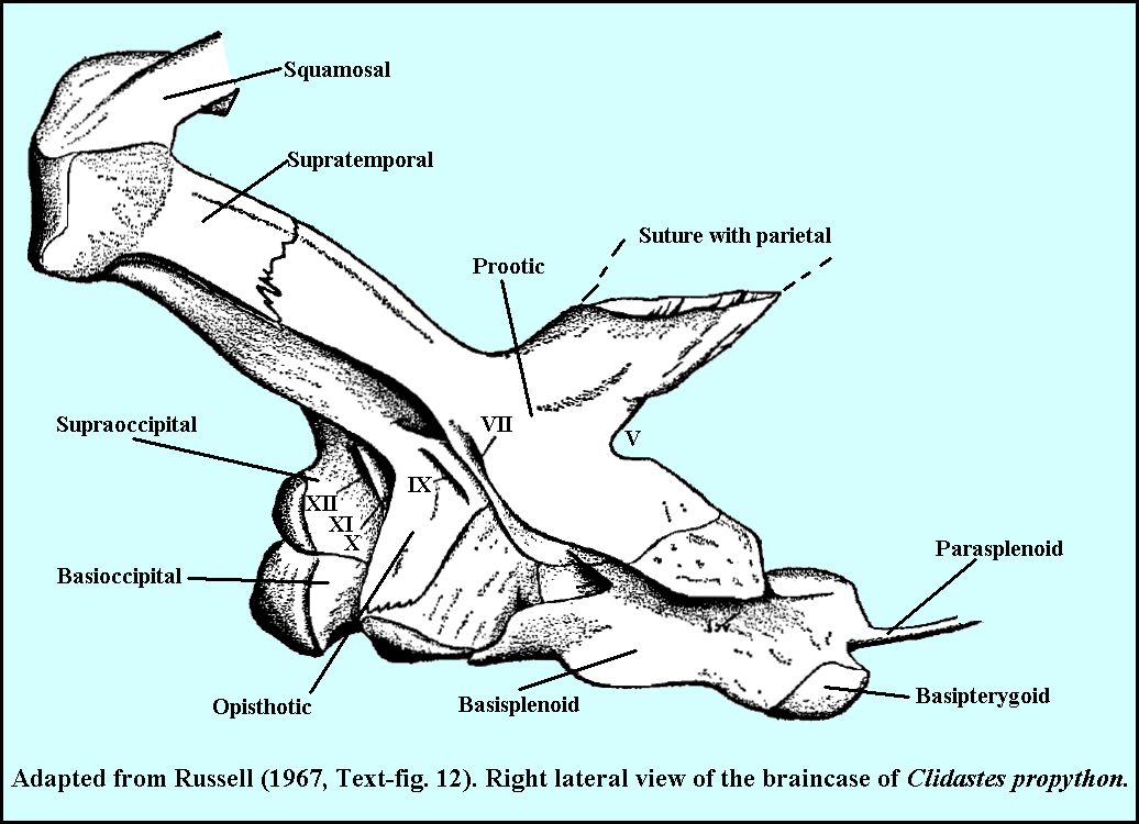
LEFT: Then we have the exterior of the braincase in
right lateral view, showing the bones that connect the braincase to the rest of the skull.
A projections from the prootic, and the paraoccipital processes of the opisthotic combine
with the surpratemporals to form two strong, strut-like bones that connect to the
squamosals to form the quadratic suspensorium... or the connection for the quadrate bone
that serves as the hinge point for the lower jaw. In addition an anterior, upward
extension of the supraoccipital meets a downward extension of the parietal along the
midline. |
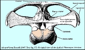 |
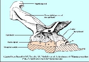
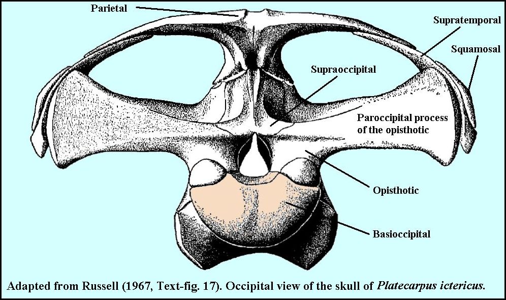
LEFT: From here, we go to the back of the skull and
show the relative position of the braincase as it is suspended within the skull: |
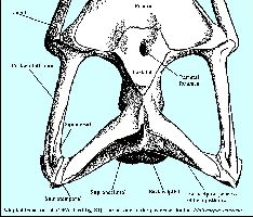 |
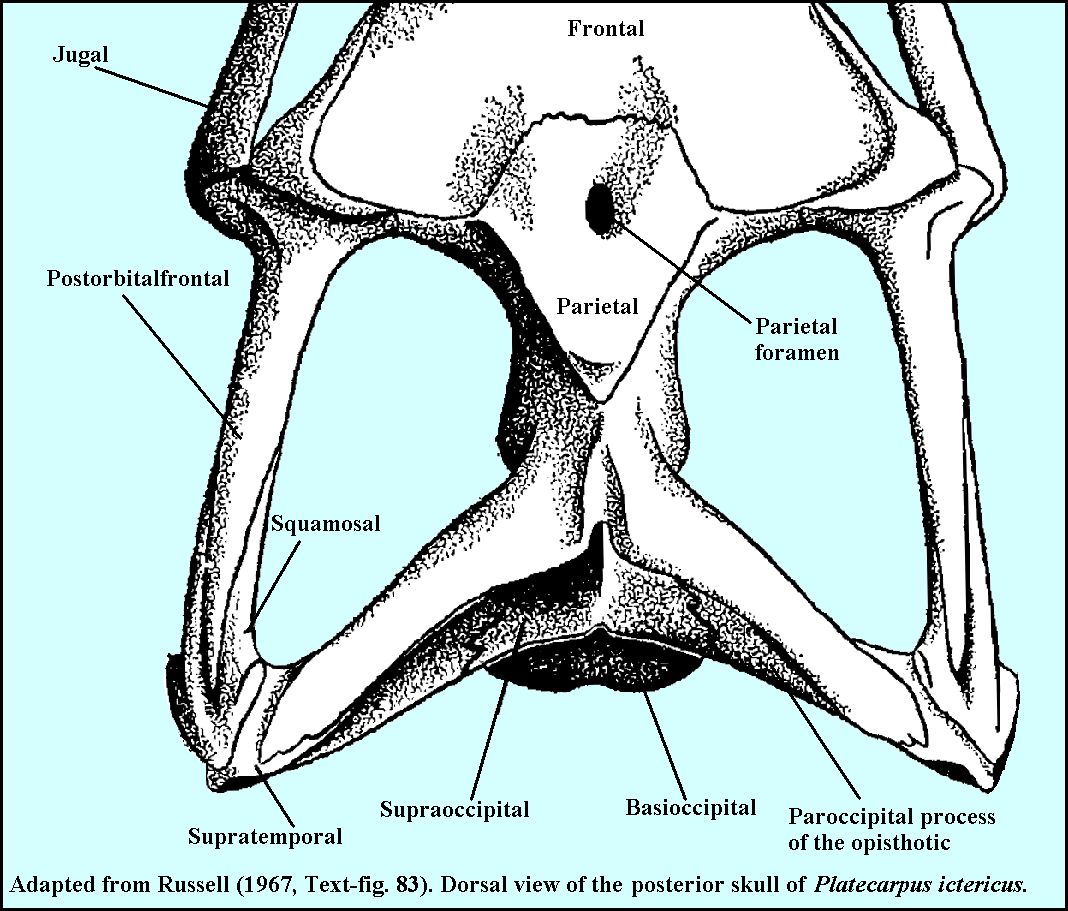
LEFT: ... then finish with a dorsal view of the
posterior portion of the skull. Note that the rearward projecting wings of the parietal
join with the paroccipital process of the opisthotic to reinforce the quadratic
suspensorium. |
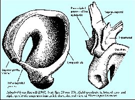 |
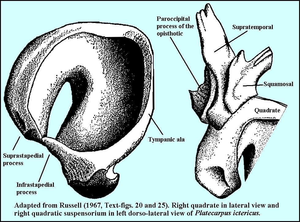
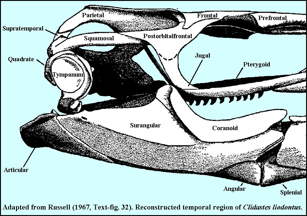
LEFT: Adding to the complexity of this part of the
skull is the somewhat flexible attachment of the quadratic suspensorium to the
quadrate. The quadrate supports the ear drum (tympanum) and also serves as the hinge
point for the lower jaw: |
 |
LEFT: The ventral portion of quadrate is the hinge
point for the lower jaw, seated in the glenoid fossa of the articular. |
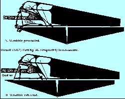 |
No discussion of the adaptations of the skull in mosasaurs is
complete without mentioning cranial kinesis and streptostyly (a special hinge joint
between the dorsal portion of the quadrate and squamosal that allows limited movement. I
will try to enlarge upon this subject in future additions to this page. In the meantime,
the reader is referred to Russell (1967) for a more detailed discussion. LEFT:
A diagrammatic view of the rotation of the quadrate in mosasaurs (From Russell, 1967),
showing the forward position of the lower jaw (protracted) and the rear position
(retracted). The position of the lower jaw is somewhat exaggerated for clarity.
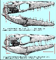
RIGHT: The skull of Platecarpus ictericus showing mouth opening, with
the lower jaw thrust forward (quadrate rocked forward) and with the lower jaw retracting
during closure (quadrate returning to a vertical position). |
 |
REFERENCES:
Camp, C. L. 1942. California Mosasaurs. University of California Press, 67 pp.
Russell, D. A., 1967. Systematics and morphology of American mosasaurs. Peabody
Museum of Natural History, Yale University, Bulletin 23.

BACK TO
OCEANS OF KANSAS PALEONTOLOGY MAIN PAGE


