A NEW MOSASAUR:
Selmasaurus johnsoni
A PHOTO GALLERY.......
Copyright © 1998-2009 by Mike Everhart
Last updated 03/26/2009
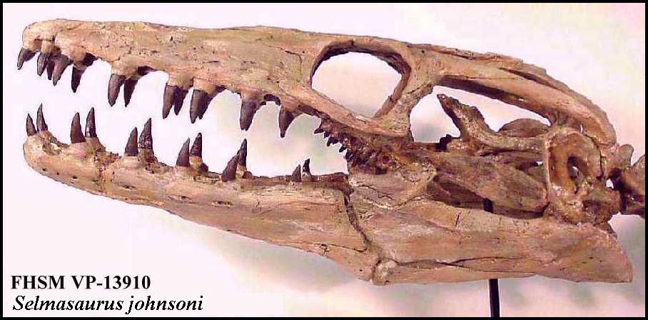
LEFT: Left lateral view of the reconstructed skull of Selmasaurus johnsoni.
|
A NEW MOSASAUR:Selmasaurus johnsoniA PHOTO GALLERY.......Copyright © 1998-2009 by Mike EverhartLast updated 03/26/2009
LEFT: Left lateral view of the reconstructed skull of Selmasaurus johnsoni. |
LEFT: A dorsal view of the major skull elements of this specimen. The front of the skull would be to the left. From left to right, the paired prefrontals flank the anterior of the frontal. Paired post-orbital frontals flank the suture between the frontal and the parietal. Along with the jugal (not shown), the prefrontal and post-orbital-frontal comprise the orbit of the mosasaur’s eye. (Scale is 10 cm) |
.
 |
The photo shows the distinctive angular shape of the frontal bone in mosasaurs. Although actually a pair of embryonic bones, they are co-ossified during development and there is no suture between them. |
 |
Dorsal (left) and ventral (right) views of the parietal. The oval opening is the parietal foramen. In P. planifrons, the foramen is set further back from the frontal/parietal suture than in other species. The mosasaur’s brain would be located beneath this bone in the rear portion of the skull. |  |
 |
LEFT: Exterior views of the left (top) and right maxillaries, showing the large teeth that appeared to be rather unusual for a mosasaur of this size. A new tooth is emerging behind the second large tooth of the right maxillary. |
 |
LEFT: A ventral view of the bones that make up the muzzle of the mosasaur (Left and right maxillaries joined by a typical Platecarpus pre-maxillary). |
 |
LEFT: A lateral view of the premaxillary and the left maxillary. The prow-like projection on the premaxillary which is visible in this photograph is a character which distinguishes this species from P. tympaniticus.
|
 |
LEFT: External views of the right and left quadrates. The quadrates are located at the back corners of the skull and serve as the hinge points for the lower jaws. They also support the tympanic membrane (eardrum) of the ear and have several distinctive characters in this species. |
 |
LEFT: The right (upper) and left (lower jaws. The flexible joint between the tooth bearing portion (dentary) and the back of the jaw is visible in this picture. |