
PTERANODONS
FLYING REPTILES OF THE LATE CRETACEOUS
WESTERN INTERIOR SEA -
A Photographic Atlas
Copyright © 2000-2013 by Mike Everhart
|
PTERANODONSFLYING REPTILES OF THE LATE CRETACEOUS WESTERN INTERIOR SEA - A Photographic Atlas Copyright © 2000-2013 by Mike Everhart Updated 12/05/2014 |
 |
Pterosaurs (flying
reptiles) were the first vertebrates to take wing and evolved during the Late Triassic.
They were superbly adapted for flight, with hollow, air-filled bones, a relatively large,
birdlike brain (Seeley, 1871; Edinger, 1927; Wellnhofer, 1991), and membranous wings that
were supported by the elongated fourth finger of each hand. In the much larger
pteranodons, their upper bodies were stiffened by rigidly binding the fused dorsal
vertebrae, ribs, scapulacoracoid and sternum together into a solid structure (notarium)
that supported the large muscles needed to power their wings. Some smaller pterosaurs were
apparently covered with “fur-like” bristles and it is likely that they were
"warm-blooded" to some extent. Besides the elongated
“wing-finger,” they had three clawed fingers
on each hand, and four clawed toes on each foot.
The smallest known pterosaur (Pterodactylus)
was about the size of an American robin, and one of the largest (Quetzalcoatlus)
had a wingspread as large as a light airplane (11-12 m/ 36-39 ft.).
Almost all of the known remains of Pteranodon come from the Smoky Hill Chalk of west Kansas. A few are found in the overlying Pierre Shale in Kansas, South Dakota and Wyoming. These are all marine deposits, as much as hundreds of miles from the nearest shoreline at the time. So what were these flying reptiles doing so far from land? The current idea is that they were feeding, but why fly that long distance just to feed? To me it seems far more likely that what we are seeing in these remains are those pterosaurs that died during migrations across the seaway, going to and from the rookeries where their young were born / hatched and raised. Modern bird migrations, especially those over water, tend to lose weak, sick or old individuals. Such losses would also occur among pterosaurs. LEFT: A flight of Pteranodon longiceps as shown in the opening scenes of the National Geographic IMAX movie, "Sea Monsters" (Released October, 2007). I was fortunate to be one of the senior science advisers on the movie, along with Ken Carpenter, Glenn Storrs, Larry Martin and others. |
| PTEROSAUR NEWS AND VIEWS:
New specimen: Pterodactyl mother found with egg in China - color photos - Lü, J., Unwin, D.M., Deeming, D.C., Jin, X., Liu, Y. and Ji, Q. 2011. An egg-adult association, gender, and reproduction in pterosaurs. Science 331(6015):321-324. (January 21, 2011) Pterosaur Blog - Updates on pterosaur discoveries around the world. Gwawinapterus - A new
pterosaur from Canada - Arbour,
V. M. and Currie, P.J.(2011. An istiodactylid pterosaur from the Upper
Cretaceous Nanaimo Group, Unfortunately it turned out to be a fish: Vullo, R., Buffetaut, E. and Everhart, M.J. 2012. Reappraisal of Gwawinapterus beardi from the Late Cretaceous of Canada: A saurodontid fish, not a pterosaur. Journal of Vertebrate Paleontology 32(5):1198-1201. Two new Pteranodon species named: Kellner,
A.W.A. 2010. Comments on the Pteranodontidae (Pterosauria, Pterodactyloidea)
with description of two new species. Anais da Academa Brasileira de Cięncias
82(4):1063-1084. |
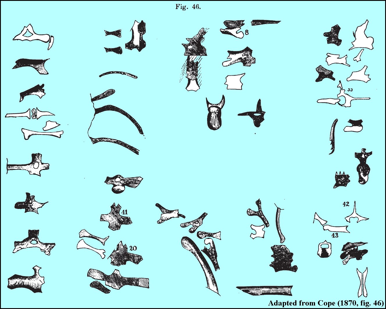 |
Cope (1866), not Marsh (1871), was actually the first
paleontologist to mention the bones of pterosaurs in North American rocks,
based on what appear to be fairly numerous small, hollow bones found in
the Triassic of Pennsylvania (see figure at LEFT from Cope, 1870) . The note is short...
just a oral presentation at the October 23, 1866 meeting of the Academy of
Natural Sciences of Philadelphia:
“Mr. Cope made a communication in regard to the Mesozoic Sandstone
of Pennsylvania, expressing the probability of its horizon being that of
the Trias of Europe, on account of some contained vertebrate remains which
he had previously described, and also from some bones of a Pterodactyle
now in his possession, for which he proposed the name of P. longispinis.” |
 |
Pteranodon
( meaning "wing without tooth") were a group of Late Cretaceous
flying reptiles that were characteristically toothless and tail-less (they did have short
tails). The males grew to large size (wingspreads of 7.5 m (25 ft) or more) during
the deposition of the Smoky Hill Chalk (87-82 mya), and were even larger near the end of
the Late Cretaceous. The
largest individuals discovered so far in Kansas had a wingspread of about 8 m (26 ft.) but
by some estimates may have weighed no more than 11 kg (25 lb). However,
in a recent article in the Journal of Vertebrate
Paleontology, Henderson (2010) estimated the mass of a Pteranodon
longiceps with a 5.34 m (17.5 ft) wingspread at 18.6 kg (41 lb).
Clearly, there is still work to be done on their anatomy and
physiology.
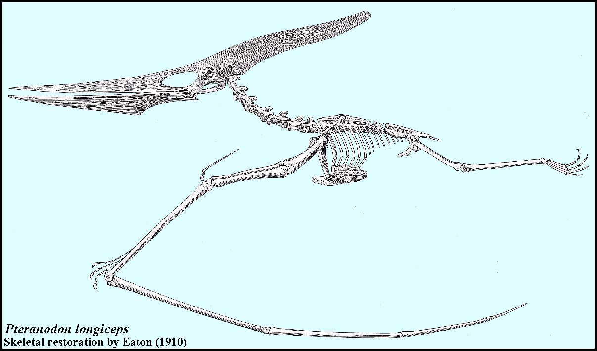
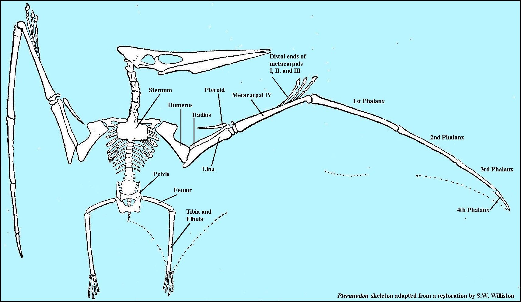
Compared to the size of their heads and wings, their bodies are almost absurdly small. One comparison provided by Hankin and Watson (1914) was that "with a body little larger than that of a cat, they had a span of wing asserted in some cases to have reached 21 feet or more!" LEFT: The skeleton of Pteranodon longiceps (lateral view) - Adapted from original drawing by Eaton (1910). CLICK FOR LARGER VERSION |
Systematic Paleontology Order Pterosauria Suborder Pterodactyloidea Superfamily Ornithocheiroidea Family Pteranodontidae Genus Pteranodon Marsh 1876 Type Species Pteranodon longiceps Marsh 1876 |
During the summer and fall of 1870, O. C. Marsh and his Yale scientific expedition collected fossils from as far west as Wyoming and Utah. Late in November, they made a brief visit to western Kansas in the vicinity of Fort Wallace where he was assigned a military escort. The weather was cold but they were still able to collect very successfully for several days along the Smoky Hill River in what are now Wallace and Logan counties. In a short note published after returning to Yale in mid-December, Marsh (1871) noted only that "some interesting reptilian and fish remains" were collected during their two-week stay in Kansas.
In his first mention of a North American pterosaur, Marsh (1871) reported that the distal ends of two long bones (two metacarpals from the wings of two individuals (YPM 1160 and 1161) had been found, and noted that they were not unlike those from Europe figured by Richard Owen in 1851. Marsh (1871, p. 472) also noted that the bones were thin-walled and hollow. From these few fragments, he named a new species Pterodactylus Oweni, "in honor of Professor Richard Owen of London." Marsh estimated the size of the creature from the fragments and noted that the outstretched wings would have measured "not less than twenty feet!" ... a very accurate estimate considering that nothing like this had ever been seen before.
Surprisingly, and as a complete fabrication since no skull material was reported to be present, Marsh also indicated that "the teeth are smooth, and compressed." In his much more complete description of additional remains of this and two other species collected by the 1871 expedition, Marsh (1872, p. 244) again noted the presence of teeth in Pteranodon: "The teeth found with remains of this species, and supposed to belong to them, are very similar to the teeth of Pterodactyls from the Cretaceous of England. They are smooth, compressed, elliptical in transverse outline, pointed at the apex and somewhat curved." Of the teeth of a second species (Pterodactylus ingens), Marsh wrote (ibid., p. 247) that "the dental characters of this species are at present only known from a single crown of a tooth; found with one series of the specimens and from two larger and very perfect teeth found by themselves, which agree so closely with the former that they deserve notice in this connection. These specimens are less curved and less compressed than the teeth referred to Pt. occidentalis, but in other respects they are nearly identical." According to Chris Bennett (pers. comm. 2003), the teeth collected by Marsh in association with his initial Pteranodon wing bones were fish teeth, probably those of an Ichthyodectes, and are still curated in the Yale Peabody collection.
Marsh, however, apparently assumed that his American "Pterodactyle" would have teeth like its European cousins and was hedging his bet on their discovery when the first skull was found. In that regard, he was not the only one who would be playing "fast and loose" with this conclusion. His rival E. D. Cope (1872, p. 337) also indicated, slightly more conservatively, that Pteranodonskulls "were slender and the teeth indicated carnivorous habits." As shown in an illustration in a book called Buffalo Land (Webb, 1872), however, it is clear that Cope believed that at least one of the species he had named (Ornithochirus umbrosus) had teeth.
In the summer of 1871, Marsh and the second Yale scientific expedition returned to Kansas. Marsh was able to return to the spot where he had found the first remains of a Pteranodon, and he located additional pieces of the same bone. By the time Marsh (1872) published more complete descriptions, however, the giant Pteranodonswere already taking a back seat to the recent discovery of toothed birds in western Kansas. Marsh noted that the name he gave to the first specimen (Pterodactylus Oweni) was preoccupied by a specimen described by Seeley and replaced it with the name Pterodactylus occidentalis. Besides this species, Marsh (1872, p. 246-247) collected several specimens of "the most gigantic of Pterosaurs," which he called P. ingens(YPM 1160 and 1172) and estimated had a wingspread "of nearly 22 feet!" Marsh also named another, smaller species (P. velox; YPM 1176) that was found in 1871 on the basis of what he believed were differences he saw in the wing bones.
Bennett (1994) examined the Marsh collection at the Yale Peabody Museum and indicated that because of the stratigraphic level in which the pterosaur remains collected by Marsh in 1871 and 1872 occurred, they were all probably Pteranodon longiceps. In that regard, Bennett (1994, p. 14) considered all the early names given by Marsh and Cope to be nomen dubia because the material does not exhibit any species-specific characters and was too fragmentary to be accurately identified beyond the genus Pteranodon.
Cope apparently found at least two sets of Pteranodonremains during his trip to Kansas in late 1871. In a short note, Cope (1872a, p. 337) named two species, Ornithochirus umbrosus (AMNH 1571) and O. harpyia (AMNH 1572), that were apparently distinguished from one another only on the basis of size. In doing so, he accepted Seeley's name for the genus and apparently ignored Marsh's Pterodactyluswithout further comment. In a narrative that preceded the listing of the two new species, Cope (ibid., p. 323) described them in their natural habitat: "The flying saurians are pretty well known from the descriptions of European authors. Our Mesozoic periods had been thought to have lacked these singular forms until Professor Marsh and the writer discovered remains of species in the Kansas chalk. Though these are not numerous, their size was formidable. One of them, Ornithochirus harpyia Cope, spread eighteen feet between the tips of its wings, while the O. umbrosus Cope, covered nearly twenty-five feet with his expanse. These strange creatures flapped their leathery wings over the waves, and often plunging, seized many an unsuspecting fish; or, soaring, at a safe distance, viewed the sports and combats of the more powerful saurians of the sea. At night-fall, we may imagine them trooping to the shore, and suspending themselves to the cliffs by the claw-bearing fingers of their wing-limbs."
While this image of pteranodons hanging from rocks along the seashore has been shown in numerous recreations over the years, it is probably just a fantasy. As noted by Stein (1975), it would have been impossible for Pteranodon to land on all fours on level ground without collapsing the wings first and thus losing lift. Performing such a landing against the vertical wall of a cliff would seem to be a death-defying act. Furthermore, none of the cores of the wing claws (unguals) I have collected or examined show any damage on the tips as might be expected from a daily routine of hanging from one's fingertips.
 |
The first Pteranodon specimens found in North America
were discovered in the Smoky Hill Chalk of western Kansas by O.C.
Marsh in 1870. The remains he collected consisted only of fragments of the long
wing bones. However, they were readily comparable (though much larger) with
pterodactyl remains from the Jurassic of Europe.
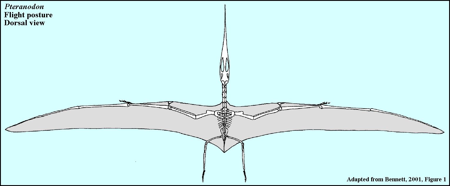
The only pterosaurs that are currently known to occur in the Smoky Hill Chalk are the highly developed pteranodons, including P. longiceps, P. sternbergi, Nyctosaurus gracilis and N. nanus. These flying reptiles do not have teeth or long tails like the earlier pterosaurs found in the Jurassic of Europe, and are generally much larger. LEFT: The current version of the skeleton of Pteranodon longiceps in flight (wingspread about 20 feet) - upper Smoky Hill Chalk, Kansas. The wing membranes are narrower than in earlier reconstructions and are no longer considered to have been connected to the lower legs. (Adapted from Bennett, 2001). This is more consistent with the wings of modern long distance flyers like the albatross and frigate bird. |
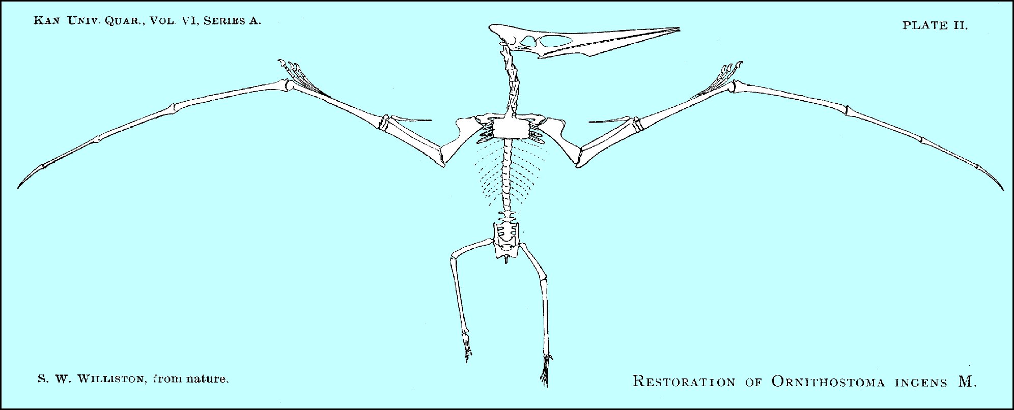 |
LEFT: One of the first drawings of a complete skeleton of a Pteranodon by S.W. Williston (1897, Plate II). Note that "Ornithostoma ingens" is a junior synonym of Pteranodon longiceps. Williston initially disagreed with Marsh on the genus name and thought that it should be the same as the pterosaur remains from England named earlier by Seeley. Williston also disagreed with Marsh in regard to the size of the crest. |
Pterosaurs were apparently "warm blooded" in some respects, and may have had a thin covering of hair on their bodies. Their role in the Late Cretaceous Inland Sea was probably similar to modern sea birds such as the albatross and pelican, and they may have spent most of their lives soaring over the ocean looking for food. While they apparently fed primarily on fish and other small marine organisms, we are not sure how they fed. It is unlikely that they were "skimmers," that is, taking fish from the ocean surface while in flight.
 |
LEFT: Another version of Eaton's 1910 drawing of Pteranodon
longiceps. In this case, the figure is an illustration from: Hankin, E. H. and D. M.
S. Watson. 1914. On the flight of Pterodactyls. The Aeronautical Journal, 18: 324-335.
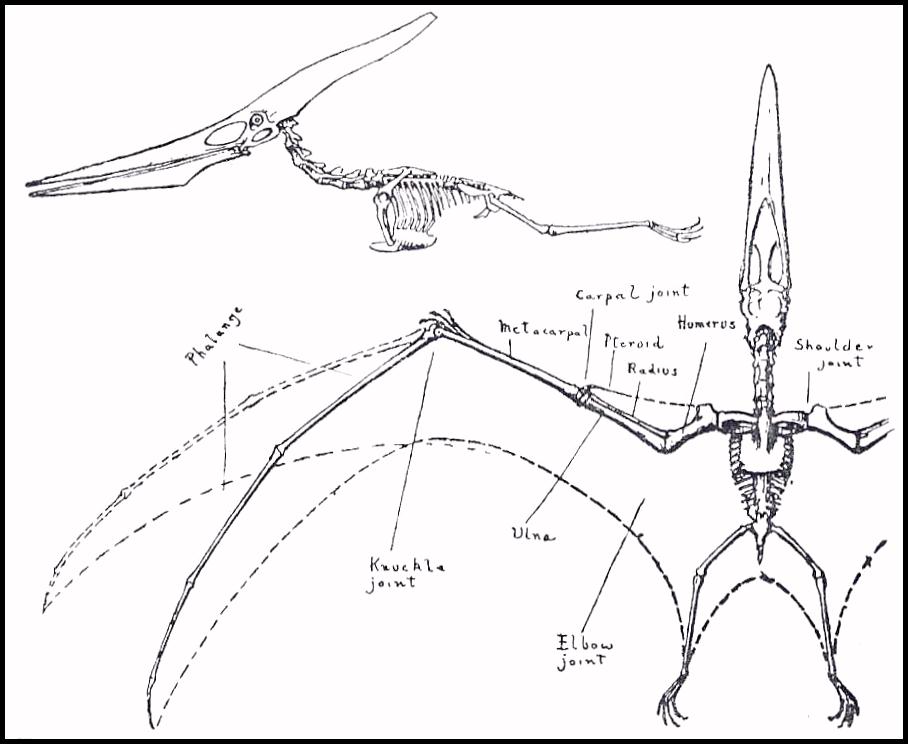
Note the small size of the Pteranodon's body compared to the length of the head, and to the wings. Pteranodons were superbly adapted to flight, including their hollow, pneumatic bones and the way in which their dorsal vertebrae were fused together with the ribs to form a solid structure (notarium) that supported the flight muscles. While they were excellent long distance flyers, they probably spent more time soaring than flapping their wings. RIGHT: Bones of a Pteranodon skeleton labeled. Adapted from a reconstruction by S.W. Williston. Note that the long wing finger corresponds to the "ring finger" in humans (Digit IV). |
 |
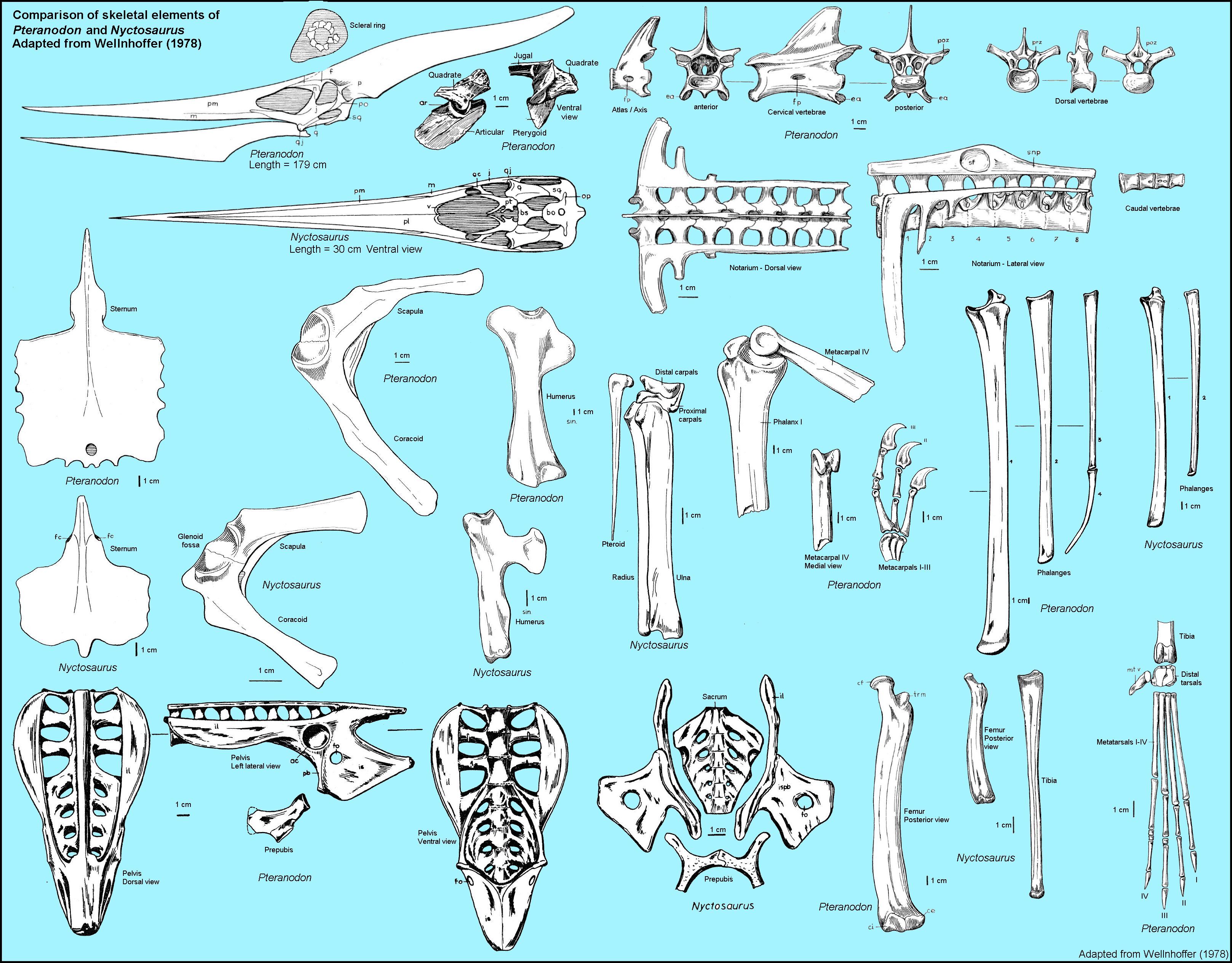 |
LEFT: A comparison of Pteranodon and Nyctosaurus skeletal elements. Note that the scales differ between bones and between genera, with Pteranodon being the larger of the two genera. Adapted from Wellnhofer (1978 - Note that most of the these images originally came from Williston's and Eaton's previous works). |
 |
These two drawings were adapted from Lucas, F. A. 1929.
Animals
of the Past. American Museum of Natural History, Handbook No. 4, New York.
LEFT: A comparison between the sizes and the skeletons of an adult Pteranodon longiceps and a California Condor - Gymnogyps californianus. The wing spread of the Pteranodon is about 24 feet, while that of the Condor is about 10 feet. RIGHT: The different bone structures of wings in bats, pterodactyls, a primitive bird (Archćopteryx) and a modern bird. |
|
Pteranodon longiceps
Pteranodons were first discovered in the upper chalk near Fort Wallace in Logan County, Kansas by O.C. Marsh in 1870. The only remains collected initially were the fragments of long wing bones, but they were recognizable as being similar to those of the Jurassic pterodactyls found in Europe. Marsh initially named two species from the remains, Pterodactylus occidentalis and P. ingens, now considered by Bennett to be nomen dubium. Pteranodon. longiceps Marsh (1876) was named from a much more complete specimen (YPM 1177) collected by S.W. Williston that included the skull. Cope also named two species from specimens that he collected in 1871 (Ornithochirus umbrosus and O. harpyia / occidentalis). It was later noted by Bennett (1994) that all of these early specimens were collected from the upper chalk and were probably from the same species, P. longiceps.
 |
At first, both Marsh (1871) and Cope (1872) contended that these giant
flying reptiles also had teeth like their Jurassic cousins. In Marsh's case, it was
because of some fish teeth that were found near the bones; Cope apparently just decided
that they probably had teeth. It wasn't until 1876 with the discovery of the skull of
the type specimen of Pteranodon longiceps (YPM 1177) by S.W. Williston in
the upper chalk of western Gove County that it was discovered that these flying reptiles did
NOT have teeth and, and subsequently the genus name, Pterodactylus, was
changed to Pteranodon by Marsh (1876).
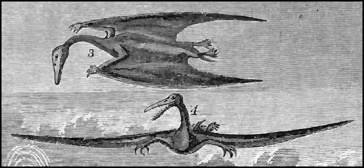
LEFT: Two reconstructions of Ornithochirus umbrosus Cope 1872 (Adapted from a plate opposite page 356 in Webb's 1872 Buffalo Land), most likely drawn from instructions provided by E.D. Cope. Note that neither pteranodon has a crest (unknown in 1872) and the lower figure (4) has teeth (assumed). |
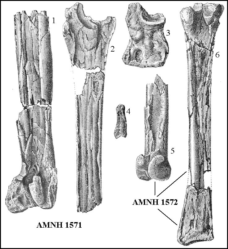 |
LEFT: Figure adapted from Plate 7 of Cope's (1875) Vertebrata of Pteranodon specimens he collected in 1871 from the chalk of western Kansas, and from which he named
two species (Ornithochirus umbrosus Cope 1872 and O. harpyi Cope 1872. Bennett (1994) identified the bones as (1) distal left metacarpal
IV and (2) proximal wing phalanx 1 of two different individuals, (3) proximal carpal and
(4) manual phalanx from the type specimen of Ornithochirus umbrosus (AMNH 1571), and; (5) distal right metacarpal IV and (6) a composite of a proximal left
wing phalanx 1 and a proximal left wing phalanx 1(?) of two individuals composing the type
specimen of O. harpyi / occidentalis (AMNH 1571).
It appears that most of the Pteranodon remains collected from the Smoky Hill Chalk and the Pierre Shale are wing bones. While these are arguably the largest and most durable in the skeleton, Hargrave (2007) suggests that they were held tightly together in life, and offered little in the way of nourishment to scavengers, similar to what is observed in modern instances of partially consumed bird remains where the wings are left more or less intact. |
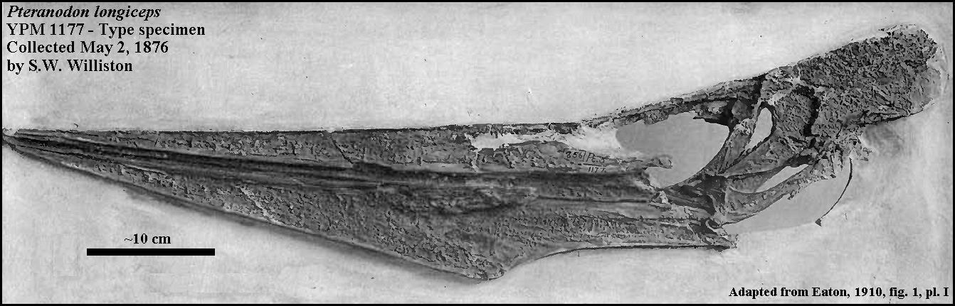 |
LEFT: The skull of the type specimen of Pteranodon longiceps (YPM 1177) in left lateral view, collected by S.W. Williston on May 2, 1876 from the
"Yellow Chalk" near the Smoky Hill River in western Kansas, and described by
Marsh (1876). Photo adapted from Plate 1 of Eaton (1910). Note that the crest is
broken off. The greatest preserved length of the skull is 73 cm (28 inches).
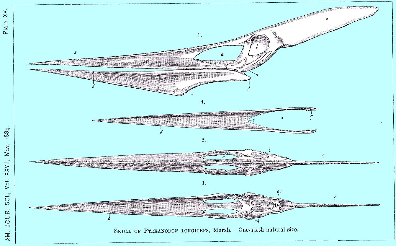
RIGHT: Plate VII from Marsh (1884) showing the skull of Pteranodon longiceps in three views. |
 |
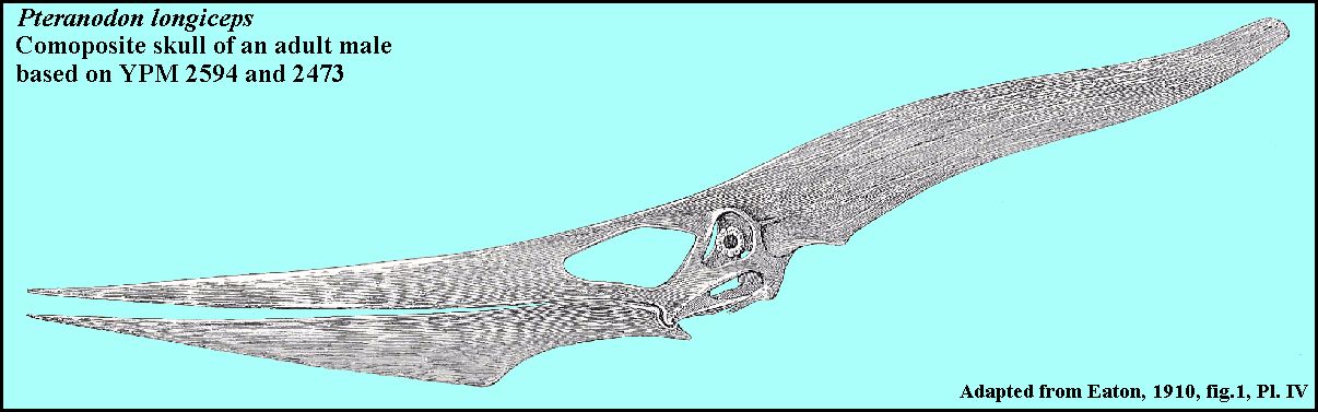 |
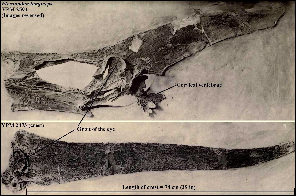
Only the mature males of Pteranodon longiceps have
the long, slender crest that extends almost as far behind the skull as the jaw extends to
the front. Females did not have a large crest.
LEFT: The reconstructed
skull of a mature male Pteranodon longiceps (left lateral view, about 4 feet).
Drawing adapted from Figure 1, Plate IV in Eaton (1910) and is a composite of YPM
2594 and YPM 2493 (a large crest) . The large crest extends about 74 cm behind the orbit
of the eye (29 inches). |
 |
 |
LEFT: A plaster model of a Pteranodon longiceps (YPM in
the Yale Peabody Museum. The exhibit is a composite based on a number of specimens
collected from the Smoky Hill Chalk, including YPM 1177 (above). It was constructed under
George Eaton's supervision for the 1904 World's Fair (Louisiana Purchase Exposition)
in St. Louis, Missouri.
RIGHT: Artist's reconstruction showing the possible development of the crests of males and females of Pteranodon longiceps and P. sternbergi. Note that although Pteranodon females do have a crest, it is never as large as those of the older, mature males. The crest apparently developed as the pterosaur reached sexual maturity, most likely as a display. Credits: This image was adapted from a slightly different version by "Smokeybjb" and shown on Wikipedia. It is displayed here with the permission of the artist. |
 |
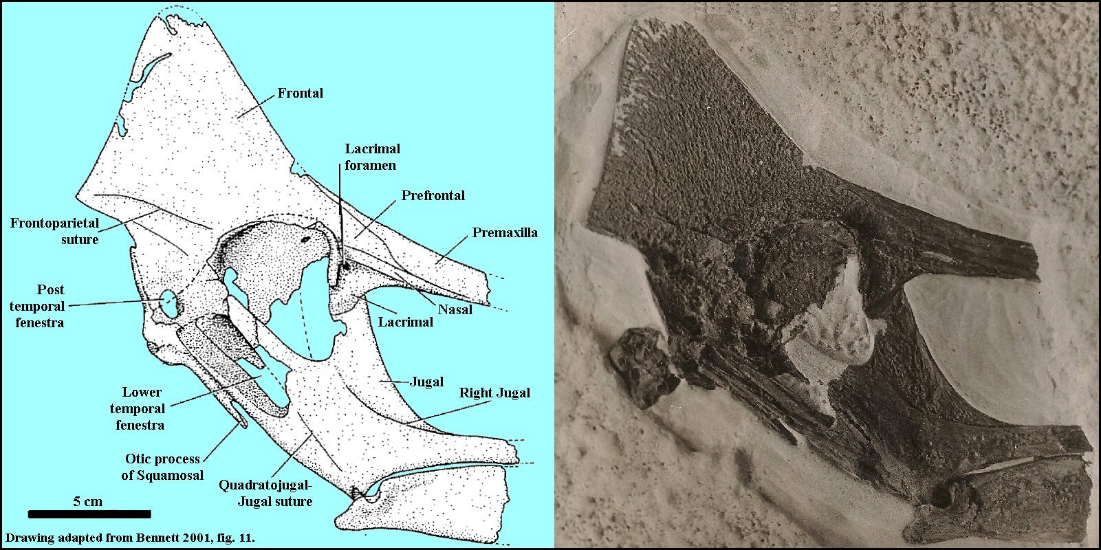 |
LEFT: A partial skull of a Pteranodon longiceps (USNM 13868) in right lateral view collected by George F. Sternberg (GFS Sp. 137-31) near Monument Rocks in western Gove County and sold to the United States National Museum (Smithsonian). Bennett (2000) suggested that it had been collected between Marker Units 15 and 16 (Upper Santonian-Lower Campanian in age). (Drawing adapted from Bennett 2001, fig. 11; Photo in the Sternberg Museum of Natural History archives). |
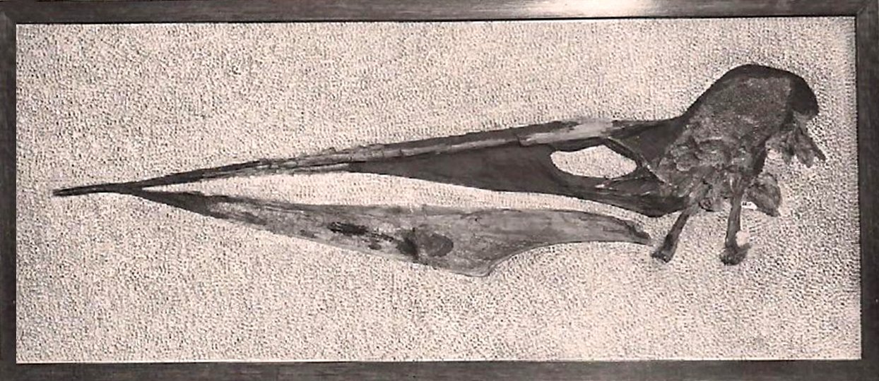 |
LEFT: The partial skull of a Pteranodon longiceps in left lateral view from the chalk of Graham County, KS. It was
collected, prepared and mounted by G.F. Sternberg. (Photo in the Sternberg Museum of Natural History archives).
Bennett (1994, p. 30) notes that the straight appearance of the upper jaw
(premaxilla) was due to the fact that the midline of the premaxilla is preserved
in dorsal view, unlike the rest of the skull.
Miller (1971, p. 15) designated this specimen as the holotype of Pteranodon (Sternbergia) walkeri. Bennett (1994) suggested that it did not differ significantly from P. longiceps. Most recently, Kellner (2010) has agreed with Bennett's (1994) re-identification. |
 |
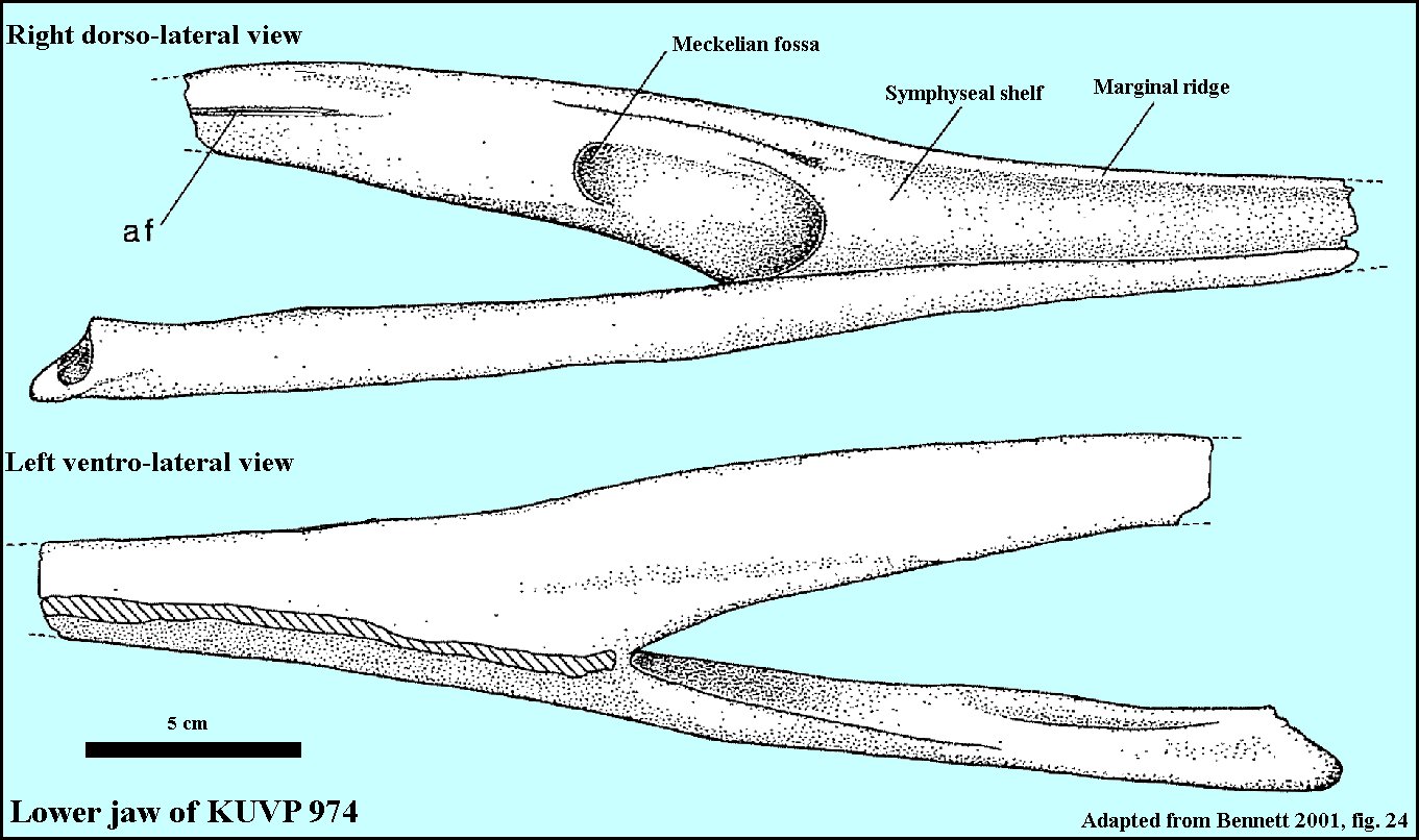
LEFT: A dorsal view of the reconstructed lower jaw of a Pteranodon,
published by Williston in 1895. The jaw is from KUVP 974 and was collected
by H.T. Martin. (Note that this figure was published upside down in
Williston's 1895 paper):
"In Fig. b is given a cross-section of the jaws at the place marked a, showing the shape of the cavity and the crushing the bone has received. At c is given a restoration of the section at a, showing the outline of jaws very nearly as they must have been during life." RIGHT: Two views of the same jaw (KUVP 974) from Bennett (2001, fig. 24). |
 |
 |
LEFT: Dorsal view
of the posterior portion of the lower jaws of AMNH 5098, collected by Charles H. Sternberg in 1877 in Lane County, showing the mass of small fish
vertebrae. Bennett (2001, pers. comm, 2004) indicated that the fish remains were probably
stomach contents regurgitated when the Pteranodon died.
RIGHT: A drawing of the jaw of AMN5098 adapted from Bennett (2001, fig. 23). Click for a photo of the complete specimen (adapted from Wellnhofer, 1991) |
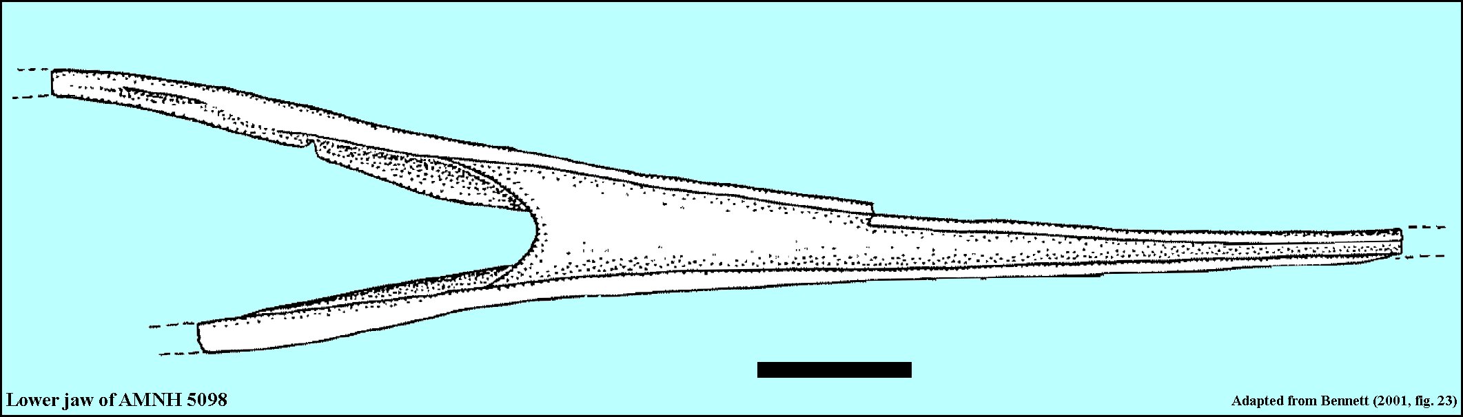 |
WHAT DID PTERANODON EAT?: It seems likely that Pteranodon fed on fish. Although not unexpected, actual evidence has been somewhat lacking. Williston (1891, p. 1126) reported finding preserved gut contents in the remains of a Pteranodon: "“Several coprolites found within the above-described pelvis, ellipsoidal in shape, and about the size of an almond, showed bones so finely comminuted that their precise character could not be made out.” These masses most likely contained the bones of small fish, but even Williston was reluctant to identify them. In his article about "Flying Reptiles" in Natural History, Barnum Brown (1943, p. 106) described the specimen shown above (AMNH 5098) and noted that it contained the "backbones of two species of fishes..." Carpenter (1996) reported a Pteranodon specimen (UCM 45062) from the Pierre Shale in Wyoming that was associated with two small coprolites containing fish bones. Hargrave (2007) reported two specimens of Pteranodon (SDSM 45719 and 69040) from the Sharon Springs Member of the Pierre Shale of South Dakota that were collected in association with fish (Enchodus) vertebrae. Beyond these two examples, direct evidence of the diet of Pteranodon has not been forthcoming.
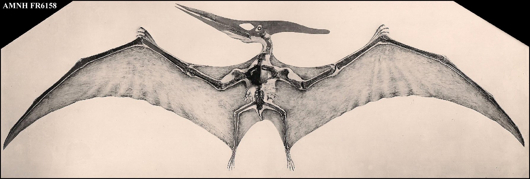 |
LEFT: AMNH FR6158 - A 1950s vintage picture of the reconstructed Pteranodon exhibit specimen at the American Museum of Natural History. The current exhibit does not include the wing
membrane. The specimen was found by H.T. Martin in 1916. It measures about 16 feet
from wingtip to wingtip.
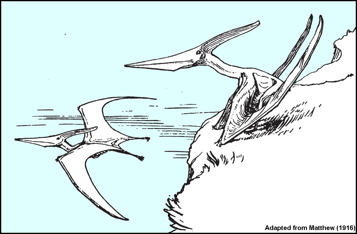
RIGHT: Two male Pteranodon longiceps take to the air in this drawing adapted from a 1916 article by W.D. Matthew in the American Museum Journal. |
 |
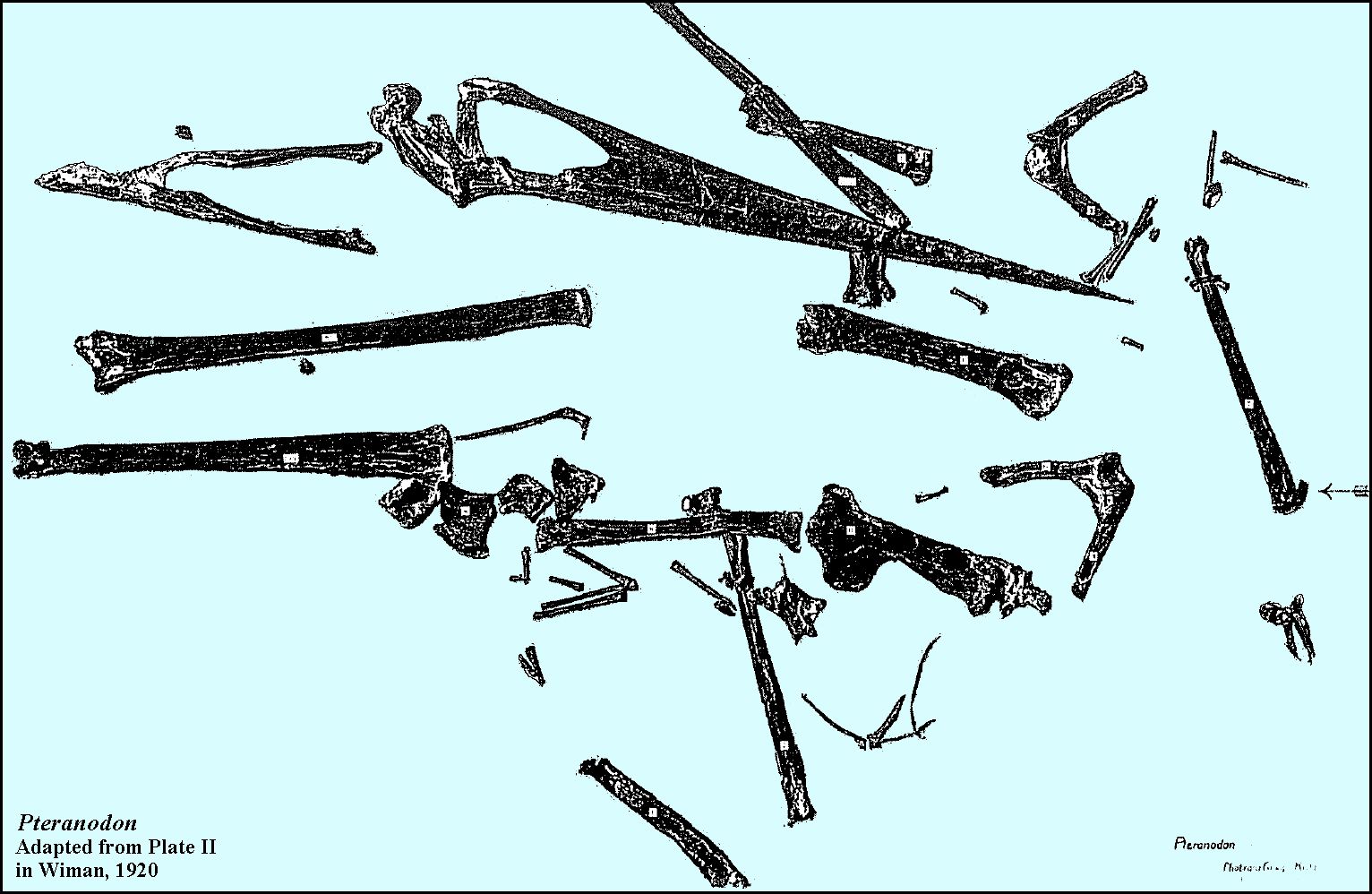 |
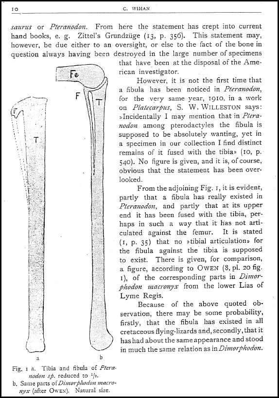
LEFT: The partial skull and skeleton of a Pteranodon collected by Charles H. Sternberg in 1918 from "12 miles south of Russell Springs in
Logan County, Kansas." The specimen was sold to Professor C. Wiman,
Upsala
University, Upsala, Sweden. The figure is adapted from a photograph of the slab mount
(Plate II) in Wiman (1920). Wiman noted that, contrary to Eaton (1910), Pteranodon does have a fibula (see arrow). Note that Williston (1891) had questioned the
presence of a fibula in Pteranodon, but later (1910) agreed that it did exist. REFERENCE:
Wiman, C. 1920. Some reptiles from the Niobrara Group in Kansas. Bulletin of the
Geological Institution of the University of Upsala, Uppsala, 18:9-18. See also: Mateer, N.J. 1975. A study of Pteranodon. Bulletin of the Geological Institutions of Uppsala 6:23-33.
RIGHT: Pages 9 and 10 from Wiman (1920). It is interesting to note that Ward's Natural History Establishment of Rochester, New York, arranged the sale of the specimen to Wiman. |
 |
 |
"TO BEAK OR NOT TO BEAK, THAT IS THE
QUESTION"
(With apologies to William Shakespeare)
Almost all other species of pterosaurs have teeth.... but Pteranodon,Nyctosaurus, Quetzalcoatlus, and a few other genera were toothless (edentulous). Did they have a horny beak like bird, such as is shown in most reconstructions?.... Good question... There is no fossil evidence for a beak, but a keratin sheath is unlikely to have been preserved except under the most favorable conditions. Here's what has been said to date on the subject:
In Marsh’s (1876, p. 1) report on the skull of Pteranodon longiceps, he says, “There are no teeth, or sockets for teeth, in any part of the upper jaws, and the premaxillary shows some indications of having been encased in a horny covering.”
However, Williston (1893, p. 2) notes that “There are no indications whatever of a horny sheath enclosing the jaw, and it is improbable that the covering of these parts was essentially different from that in the slender jawed Pterodactylidć. In texture, the maxillaries are fine-grained, and wholly without the vascular foramina found in the corresponding bones of birds. The bones are composed of two thin and firm plates, separated by cavities which are bounded by irregular walls of bony tissue. In compression from which all Pterodactyl bones have suffered more or less, the greater resistance of those walls has cased irregularities upon the outer and inner surfaces. At the borders of the bones, where the thickness has been greater, the roughening is not observed.”
Eaton (1910, p. 3) states that “The margins of the jaws are smooth and thin, though not especially sharp, and no remains of horn sheaths have been observed.”
Bennett (2001, p. 10) notes that “The marginal ridges of the upper and lower jaws appear to have aligned with one another to provide a firm grip on prey items. The marginal ridges were probably covered by a horny sheath, but there is no direct evidence of this. There are no sulci for blood vessels such as are seen in the jaws of birds, turtles and some pterosaurs (e.g., Dsungaripterus, Young 1964, fig. 2). He also adds (2001, p. 32) that in one specimen (UNC 7), "the tip of the dentary is preserved and tapers to a point less than 1 mm in diameter."
All seem to agree that the bony tips of the upper and lower jaws extended to a sharp point... but no one seems to know what, if anything, that may have covered the bone besides a layer of epithelial tissue and possibly a thin layer of keratin. This seems like a rather fragile arrangement for a large animal, especially in view of the fact that few, if any injuries (pathologies) have been observed in tips of Pteranodonjaws. It certainly casts a doubt on Pteranodon "skim feeding" as they flew over the surface of the ocean. So, how were they feeding?
At this point, the jury is still out... and the question (above) remains open.
| Although I had collected scraps of Pteranodon bones on several occasions in the late 1980s, none of these specimens were readily identifiable. In 1990, however, things started to change. |
 |
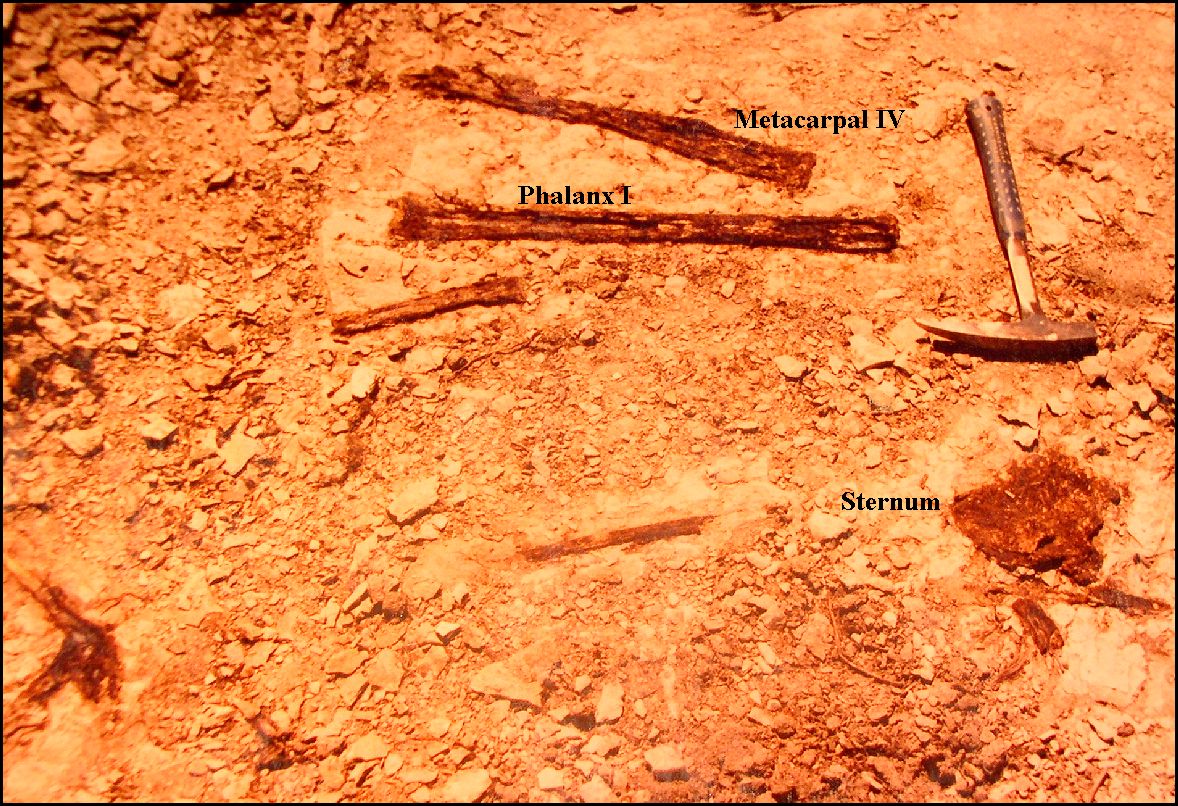
LEFT: In May, 1990, I came across some bone fragments eroding from
the chalk in Lane County. After clearing away some of the over burden, I found a complete
lower jaw of a Pteranodon sternbergi setting upright in the chalk. It had been
covered with plant roots and is extremely fragile, but I managed to recover it intact.
This was my specimen EPC1990-40. It has been donated to the Sternberg
Museum and is now FHSM VP-17702.
RIGHT: The following month I went back to the site and continued the dig, finding the sternum and the bones of a more or less complete wing. Click here for a closer view of the metacarpal IV and phalanx I. The specimen also included the sternum, vertebrae, and some leg bones. Based on the work done by Bennett (1992), I determined that this individual would have had a wing span of 4.7 m and was probably a sub-adult male. |
 |
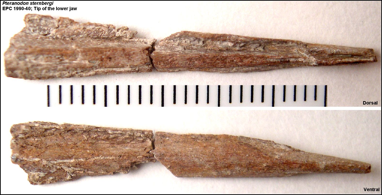 |
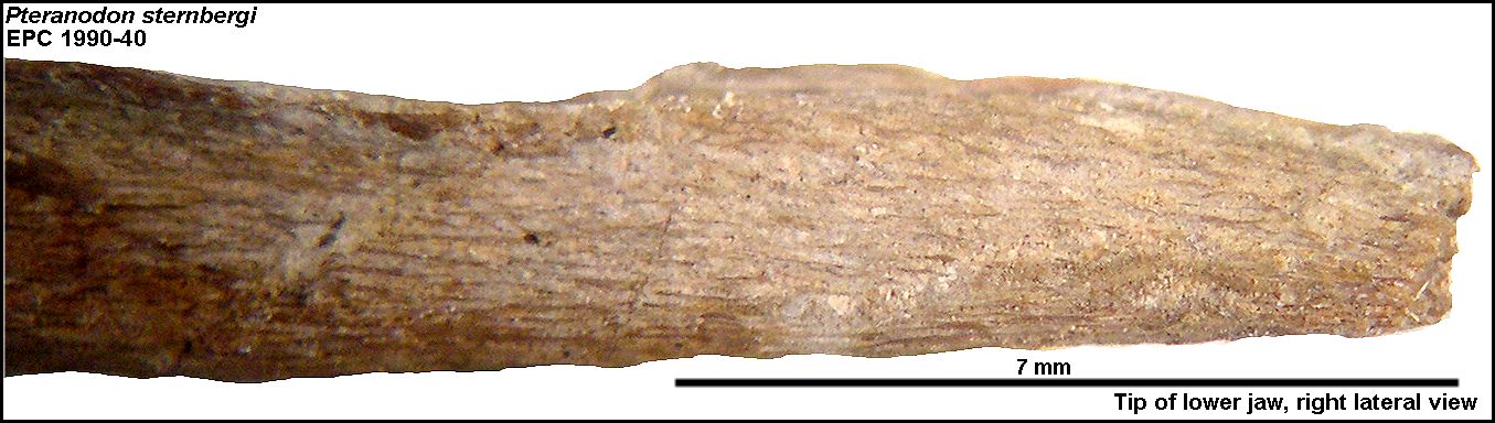
LEFT: The tip of the lower jaw of FHSM VP-17702 in dorsal and
ventral view (Scale = mm). It only took me 19 years to get around to cleaning it! The bone was covered with a mixture of chalk,
roots and preservative. Surprisingly, this little piece of bone is pretty tough and
was reasonably easy to prepare under a dissecting scope. So far as I am aware, there are
no other pictures of the tip of a Pteranodon lower jaw anywhere near this scale.
RIGHT: The extreme tip of the lower jaw in lateral view. Although it appears to go to a sharp point in dorsal and ventral view, it actually flattens out to a straight, "screwdriver"-like edge in lateral view. |
 |
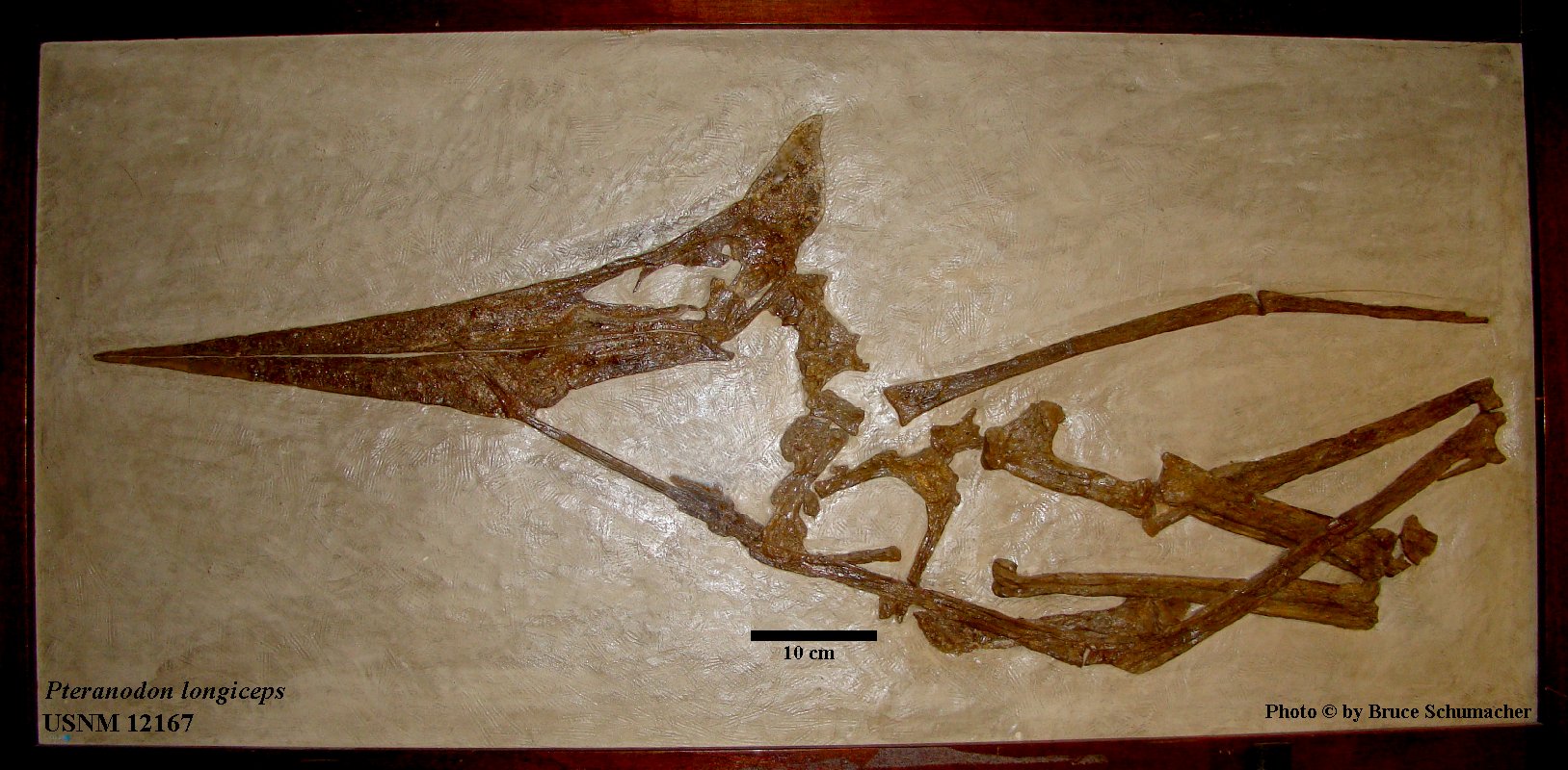 |
LEFT: A very nice Pteranodon skull and part of the
post-cranial skeleton (USNM 12167) collected by G.F. Sternberg from southeast of Elkader
in western Gove County in early 1931 (GFS 9-31). His description reads: "A nearly complete skull with lower jaws in place. Right side up,
the end of both beaks about three inches were destroyed by plant roots and cracks; the
crest is very fragile but almost all present. Length of the skull from back of the crest
to tips of beaks two feet and five inches. From back of the crest to where the distal end
is broken off is two feet and two inches. There are
seven large vertebrae which leave the skull and lay somewhat scattered, then there seems
to be several smaller vertebrae. There are wing and limb bones. Parts of the pelvic arch
and a number of broken and damaged small bones likely the feet."
The scale (10cm) is approximate but is based on data published by Bennett (2001) for this specimen. The length of the humerus ('L' shaped bone at center) is 168 mm. According to Bennett (1992), this would be near the average size of a female Pteranodon humerus. The specimen is currently on exhibit in the Smithsonian (USNM) in Washington, D.C. Another photo here |
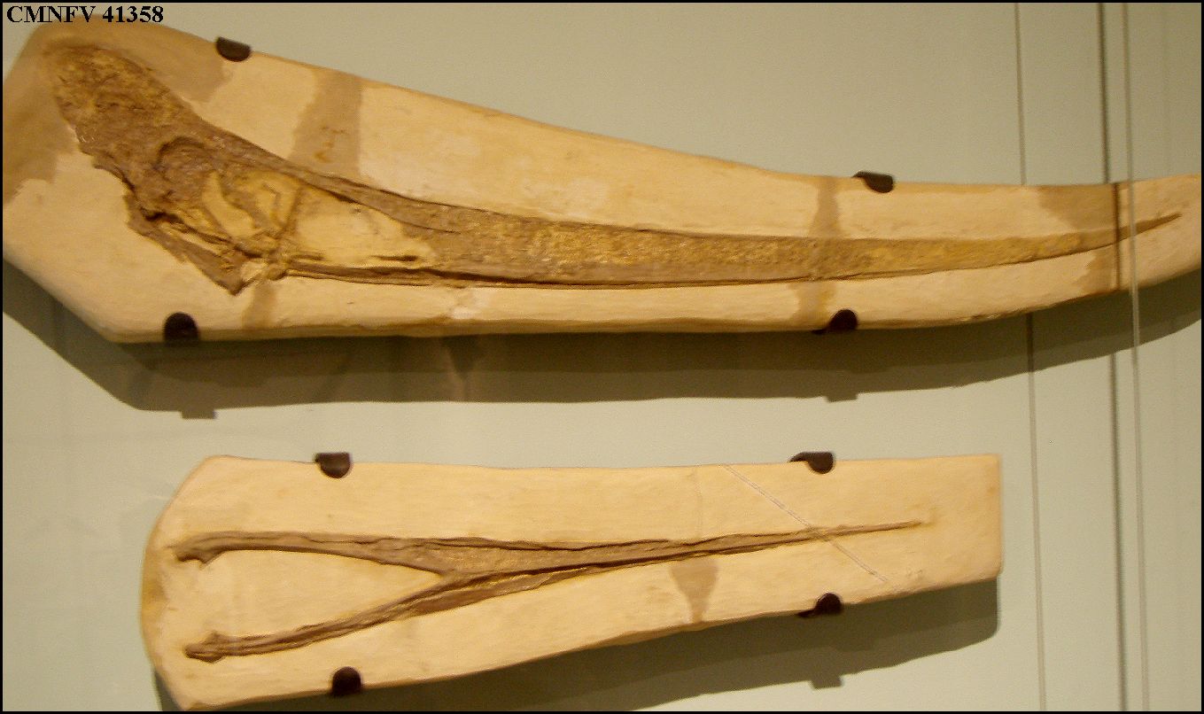 |
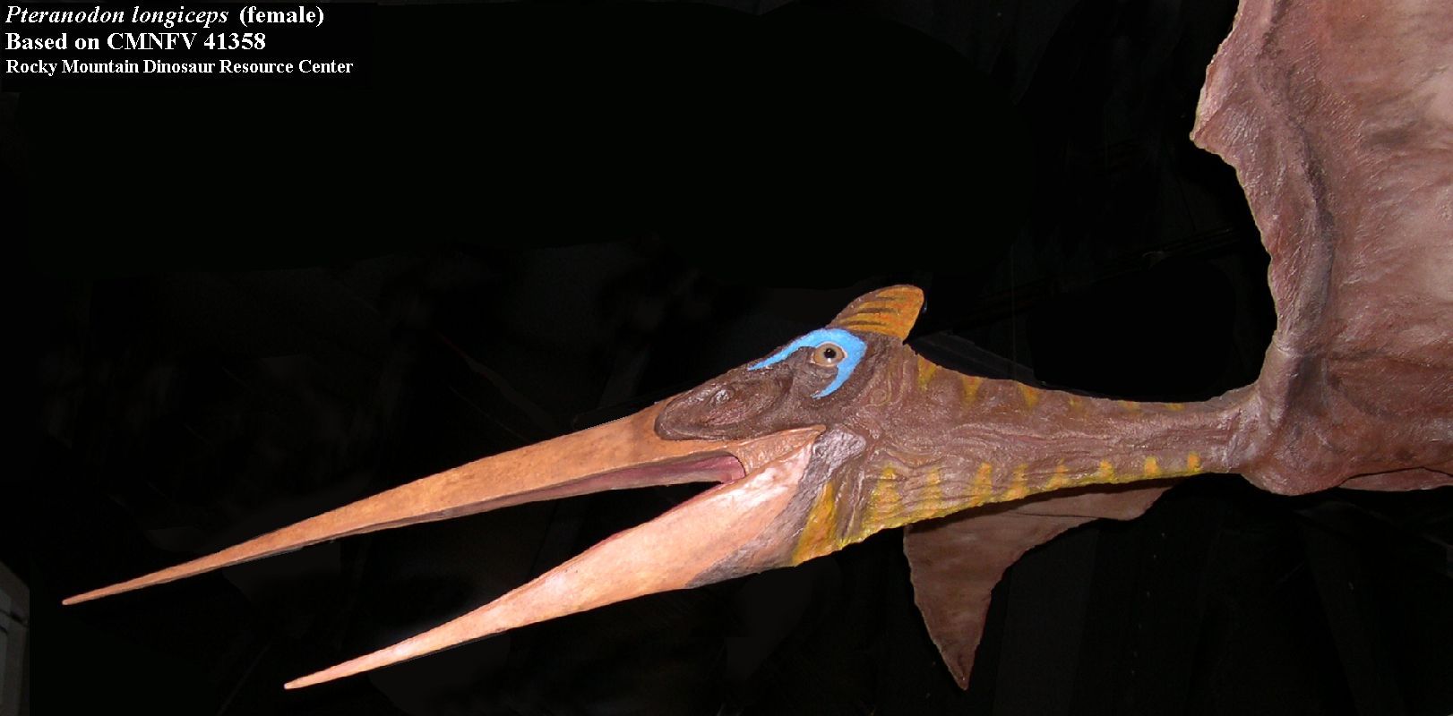
LEFT: The skull and lower jaws of a female Pteranodon
sternbergi (CMNFV 41358) on display in the Canadian Museum of Nature, Ottawa, Canada.
It was collected by Mike Triebold in 1991 from Lane County, Kansas. It is the most
complete known Pteranodon skeleton and serves as the basis for various museum
displays, like this one in the Denver Museum
of Nature and Science. Wingspan is 11 feet (3.35 meters). Photo by Steve Cumbaa.
RIGHT: A life-reconstruction based on the same skeleton (CMNFV 41358) as displayed in the Rocky Mountain Dinosaur Resource Center, Woodland Park, Colorado. |
 |
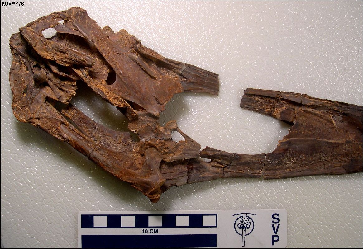 |
LEFT: The posterior portion of a Pteranodon skull (KUVP
2216 (old 976)) in right oblique view collected in Logan County by Charles H. Sternberg. This well preserved specimen is only partially
crushed and shows the palate as well as the occipital condyle in this view. Note that
the right quadrate and the quadratojugal are missing, but that the left quadratojugal and
condyloid process are complete.
RIGHT: The skull of KUVP 2216 in left oblique view showing the location of the orbit for the left eye and the base of the broken crest. A reconstruction of this skull was published by Williston: Williston, S. W. 1896. On the skull of Ornithostoma. Kansas University Quarterly 4(4):195-197, pl. I. |
 |
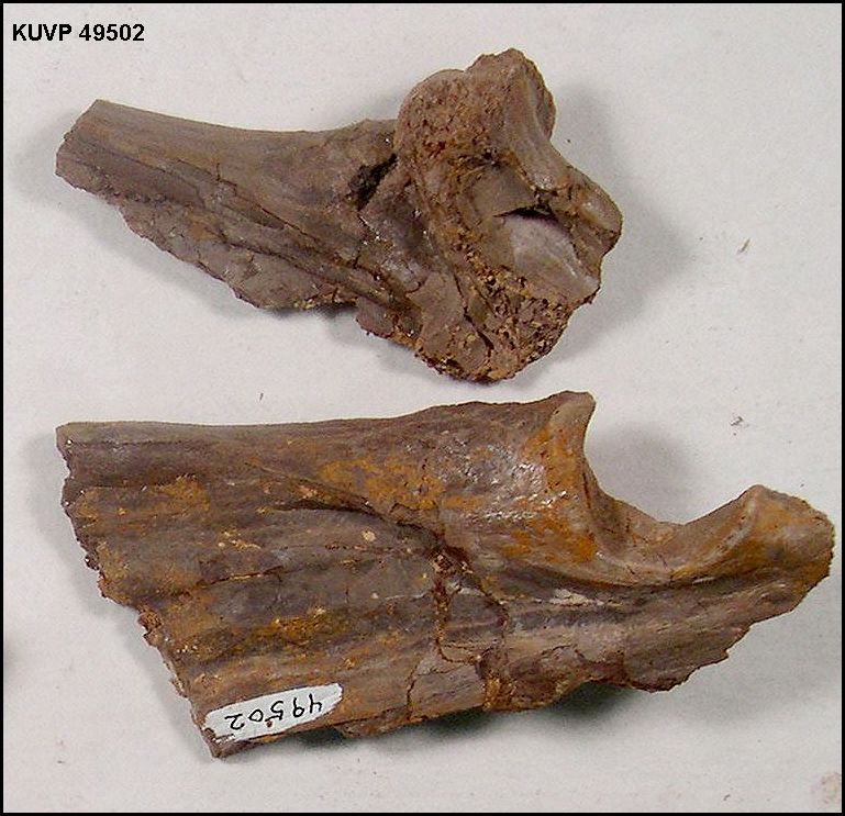 |
LEFT: KUVP 49502 - Distal end of the quadratojugal, and posterior
end of the lower jaw of a Pteranodon, including the semi-circular glenoid
process..
RIGHT: Close-up of the hinge joint of the lower jaw at the back of the skull in Pteranodon (KUVP 924) showing the quadratojugal of the skull and the articular of the lower jaw. |
 |
Post-cranial Skeleton
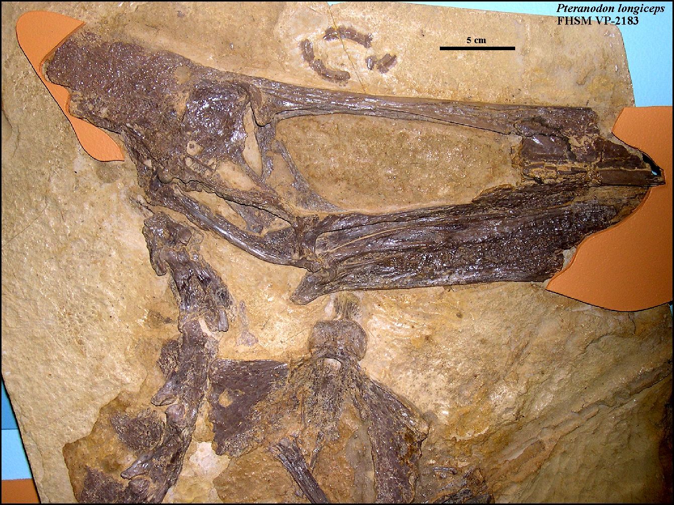 |
LEFT: Partial skull, cervical vertebrae and sternum of a female Pteranodon
longiceps (FHSM VP-2183) in right lateral view. Note the small bones of the scleral
ring above the skull. This fairly complete specimen was collected by G.F. Sternberg
near Elkader in Logan County, and described by H.W. Miller in 1971. The specimen was
somewhat unique in that it was the first P. longiceps to be described from both
cranial and post-cranial material (Marsh's holotype - YPM 1177 was a partial skull and
other fragments).
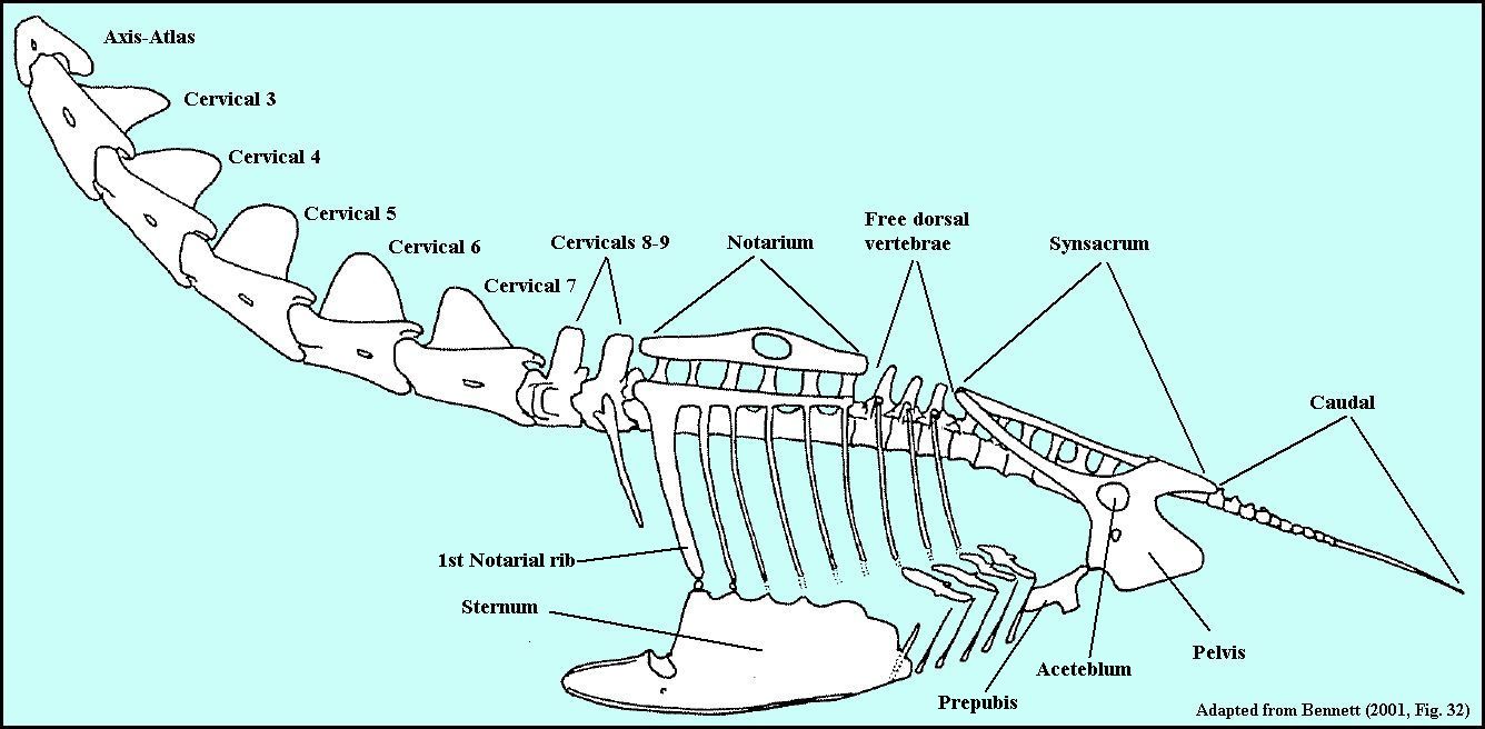
RIGHT: Axial skeleton of Pteranodon in left lateral view (Adapted from Bennett, 2001, fig. 32). Note that while the neck is fairly flexible, the most of the rest of the vertebrae are fused together into the notarium and the synsacrum to provide a solid base for anchoring the wings and wing muscles. For clarity, the scapulo-coracoids are not shown in this figure. As reported recently by Claessens, et al. (2009), pterosaurs used a "skeletal breathing pump" in their highly efficient respiratory system. |
 |
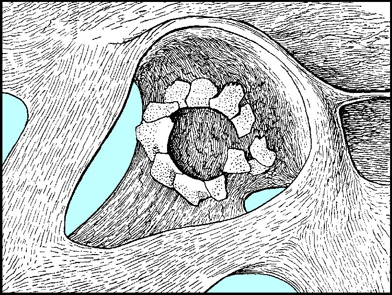 |
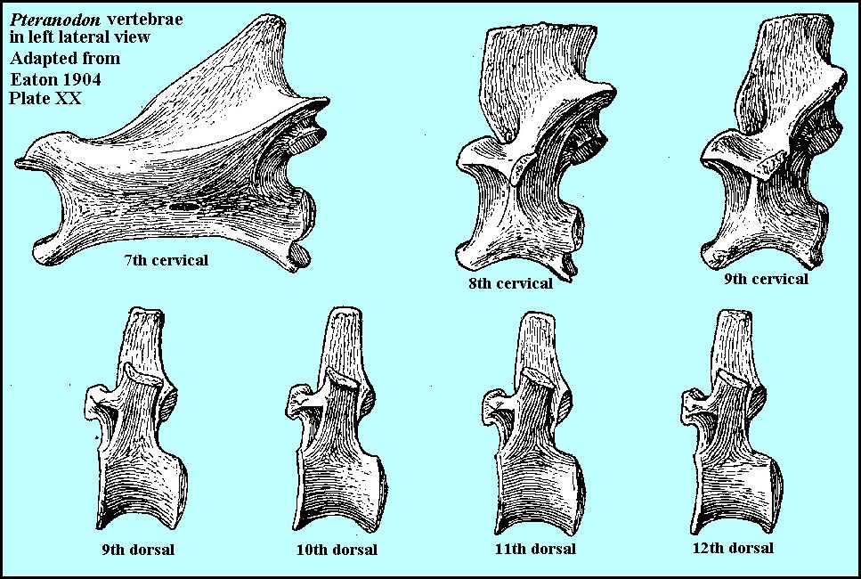
LEFT: The left scleral ring of a large Pteranodon specimen in the Yale Peabody collection as figured described by Eaton (1904, Pl. XX). The
skull is facing to the left.
RIGHT: Pteranodon vertebrae in left lateral view as figured and described by Eaton (1904, Pl. XX). |
 |
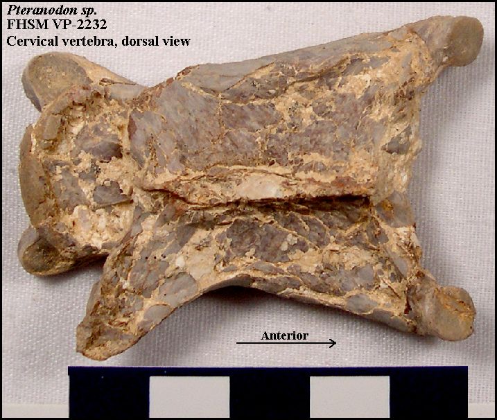 |
LEFT: A crushed Pteranodon mid-cervical vertebra (FHSM
VP-2232) in dorsal view, anterior (head) to the right.
RIGHT: Drawing of a Pteranodon mid-cervical vertebra in dorsal view with labels (adapted from Bennett, 2001). |
 |
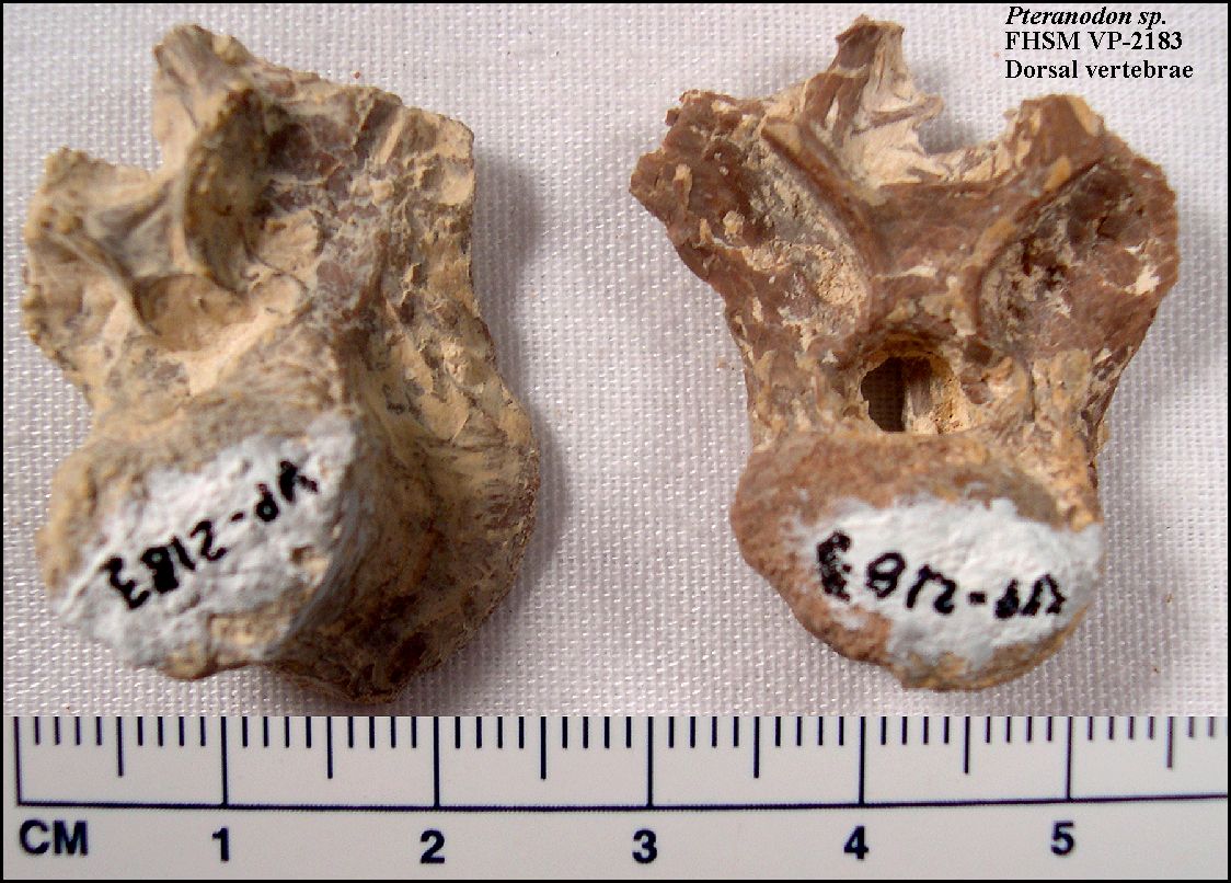 |
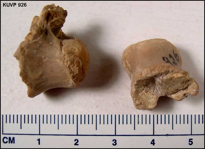
LEFT: Dorsal(?) vertebrae in posterior view from the FHSM VP-2183
specimen. Pteranodon had 9 cervical, 12 dorsal, 6
sacral and at least 11 caudal vertebrae (Bennett 2001). Like other bones of Pteranodon,
the vertebrae are usually found crushed.
RIGHT: Two dorsal vertebrae from KUVP 926, |
 |
 |
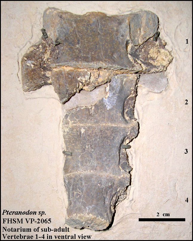
LEFT: Base of the neck (cervical vertebrae 7, 8, and 9) and the
first 3 vertebrae of the notarium of KUVP 27828. The structures jutting out at an angle
from either side are the 1st notarial (and largest) ribs. Note that this specimen also
includes a lower jaw as figured by Bennett (2001, Fig. 25).
RIGHT: The first 4 vertebrae of the notarium of an immature Pteranodon in ventral view (FHSM VP-2065). This specimen was collected by G.F. Sternberg from Logan County in 1947. |
 |
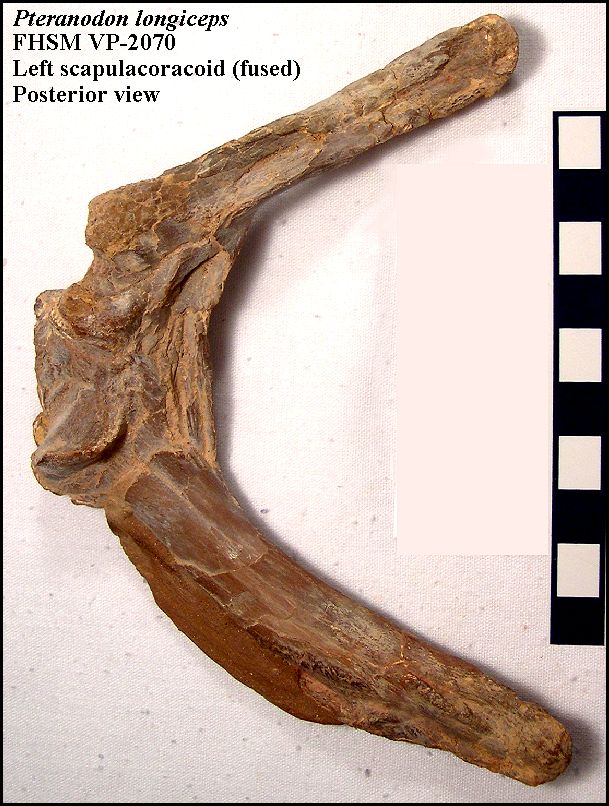 |
LEFT: The left scapulocoracoid of FHSM VP-2070 as viewed from
behind. This bone represents the fusion of the scapula and coracoid. The scapula portion
is attached to the vertebral column and the coracoid portion is attached to the sternum to
form a strong structural support for the wings. Specimen collected by M.C. Bonner in 1958
south of Russell Springs in Logan County. The right
scapulocoracoid of FHSM VP-2142 as viewed from the front. (Specimen collected by G.B.
Pierce in 1950 from near Gove in Gove County).
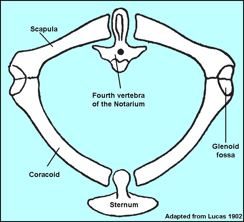
RIGHT: An anterior view of the pectoral girdle of Pteranodon, adapted from Lucas (1902). So far as I am aware, pterosaurs are the only vertebrate group where the scapula is actually attached to the vertebral column. |
 |
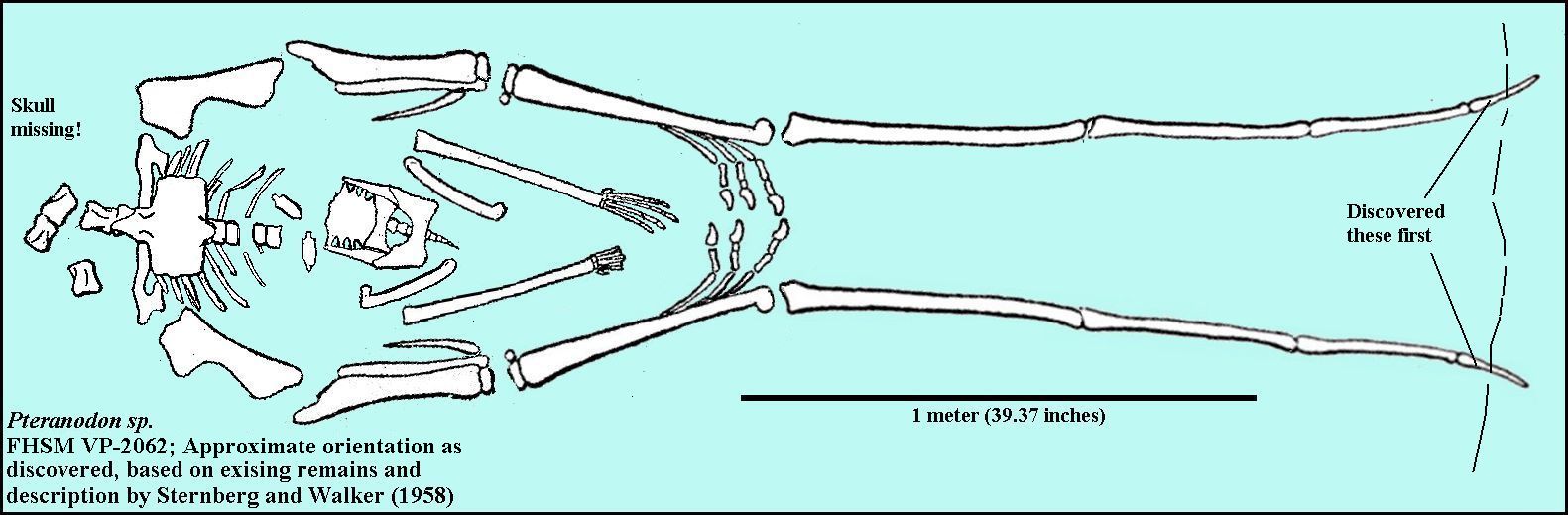 |
LEFT: My interpretation of the approximate orientation of the bones in the headless skeleton of FHSM VP-2062, based on the comments of G. F. Sternberg (Sternberg and Walker, 1958) and the existing remains. The tips of both wings (wing phalanx 4) were discovered eroding from the chalk of northeast Trego County. Both the wings and legs were still mostly articulated, while the axial skeleton was scattered and incomplete. As shown below, the claws of the wing fingers were almost touching. (See labeled Pteranodon above for identification of bones) |
 |
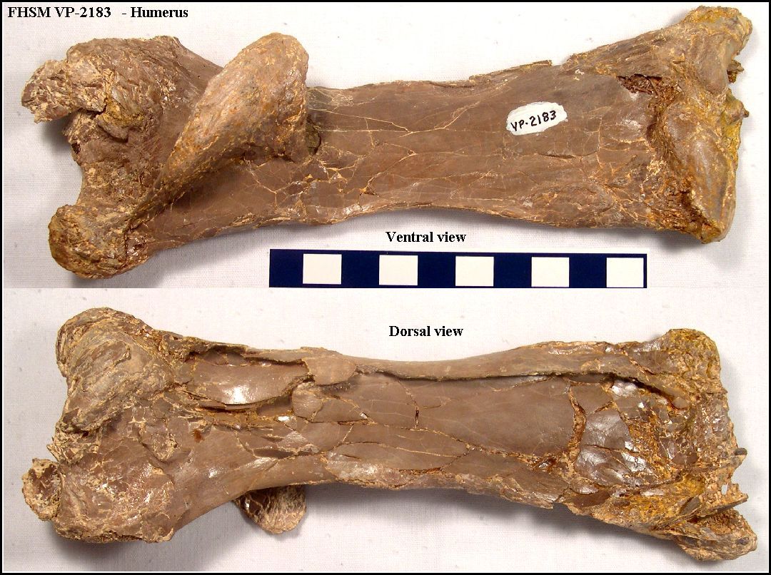
LEFT: The left (lower - ventral view) and right (upper - dorsal
view) humeri of FHSM VP-2183. The length of these bones is 18.2 cm.
RIGHT: Two views (ventral and dorsal) of the left humerus recovered with the FHSM VP-2183 specimen. Length = 18.2 cm. |
 |
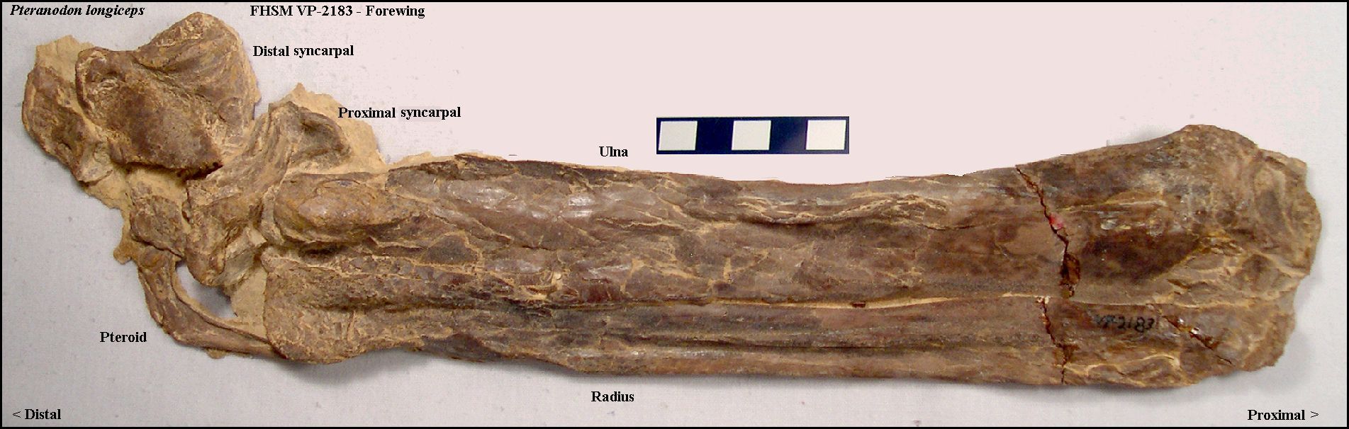 |
LEFT: The radius, ulna and pteroid bones of FHSM VP-2183. The pteroid bone was once considered to be the 'thumb' (e.g. Digit I) in pterosaurs. The two other pieces of bone in the upper left of the photo are the proximal and distal syncarpals (wrist bones) that would connect to metacarpal IV. Specimen collected by G.F. Sternberg in 1958 near WaKeeney in Trego County. Another specimen (FHSM VP-2072) is shown here in the plaster jacket used to recover it in the field. |
 |
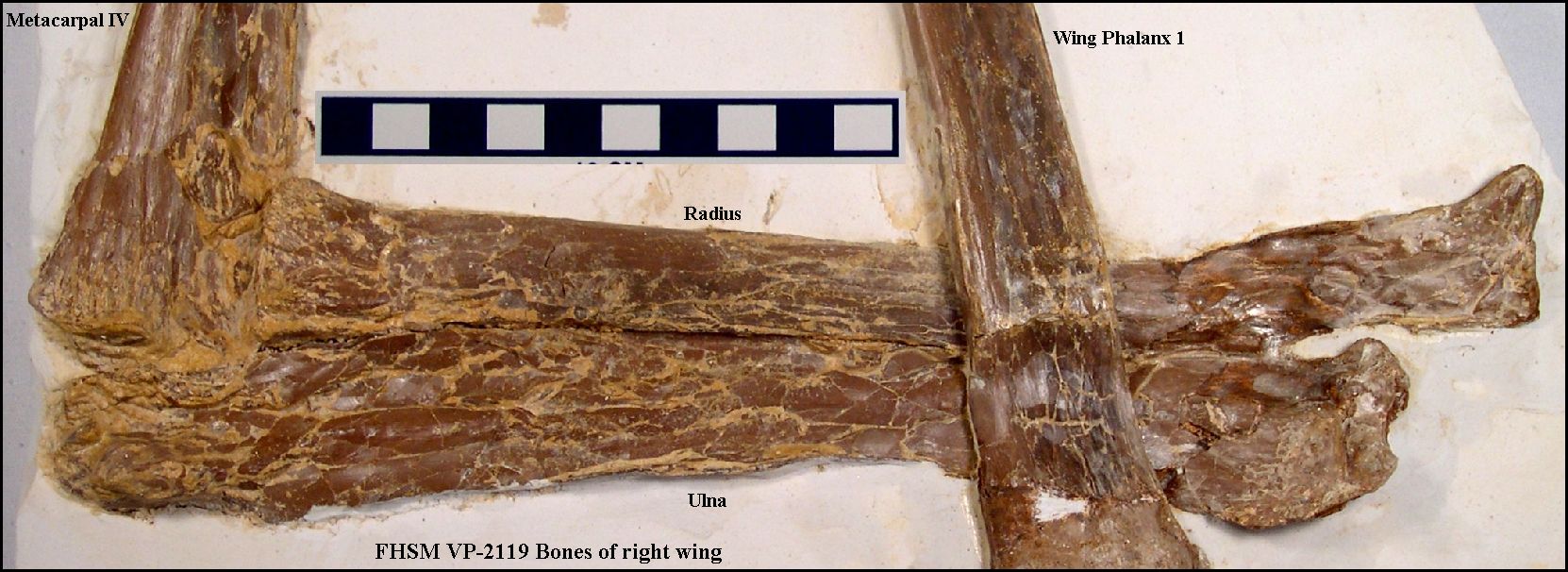
LEFT: The articulated mid-wing of FHSM VP-2119, showing the radius
and ulna, metacarpal IV and the 1st wing phalanx. Note that the proximal and distal
syncarpals are missing from this specimen. According to data from Bennett (2001, Table
11), metacarpal IV is the most commonly preserved bone of Pteranodon.
RIGHT: A closer view of FHSM VP-2119, showing the radius and ulna. Specimen collected by Marion and Orville Bonner in Logan County. |
 |
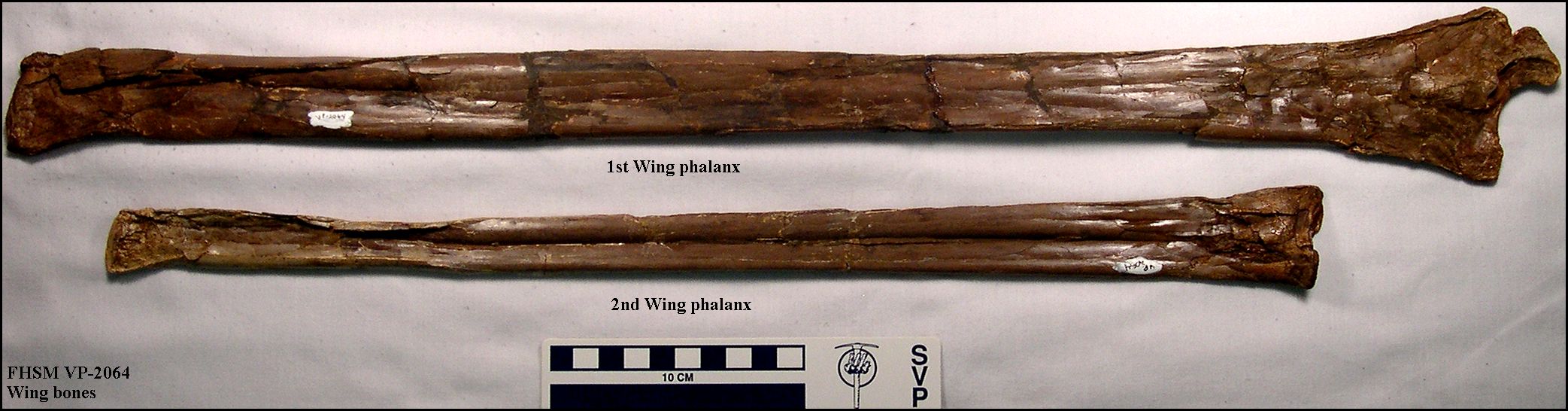 |
LEFT: The very large 1st wing phalanx (top) and 2nd wing phalanx
of FHSM VP-2064. The first wing phalanx is the longest bone in the pteranodon's wing.
Collected by G.F. Sternberg in 1953 near Moreland, Kansas (Graham County). The length of
the 1st phalanx is 60.6 cm and the length of the 2nd phalanx is 48.5 cm.
Note that according to Bennett (2001), another FHSM specimen (VP-184) is actually larger (65.3 cm and 54.9 cm, respectively; collected by G.F. Sternberg in 1949)... but the record holder at this point is probably the YPM 2833 specimen... WP1 = 69.2 cm; WP2 = 54.5 cm (est.). Figures provided by Bennett (1994) suggest that the length of WP1 is about 11% of the total wingspan of a Pteranodon. Using that figure, the wingspan of YPM 2833 would be about 7.6 m or just short of 25 feet! A very large male.... |
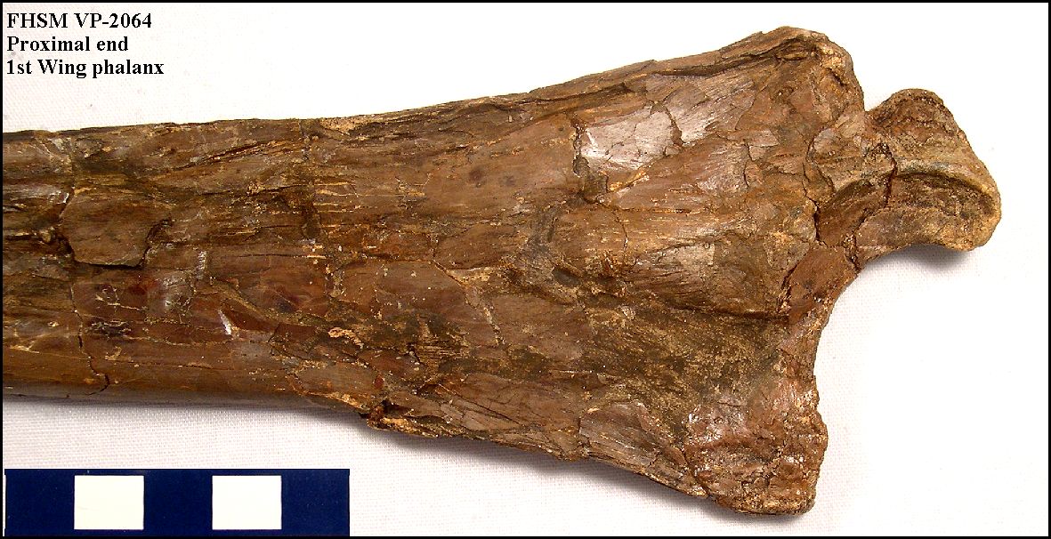 |
LEFT: A close-up view of the proximal end of the 1st wing phalanx
(above) of FHSM VP-2064.
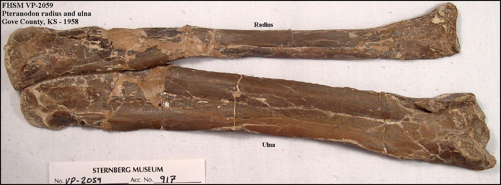
RIGHT: The radius (upper) and ulna of the wing of FHSM VP-2059. Specimen collected by G.F. Sternberg in 1958 from Gove County. Radius length = 20.5 cm; Ulna length - 21.1 cm. |
 |
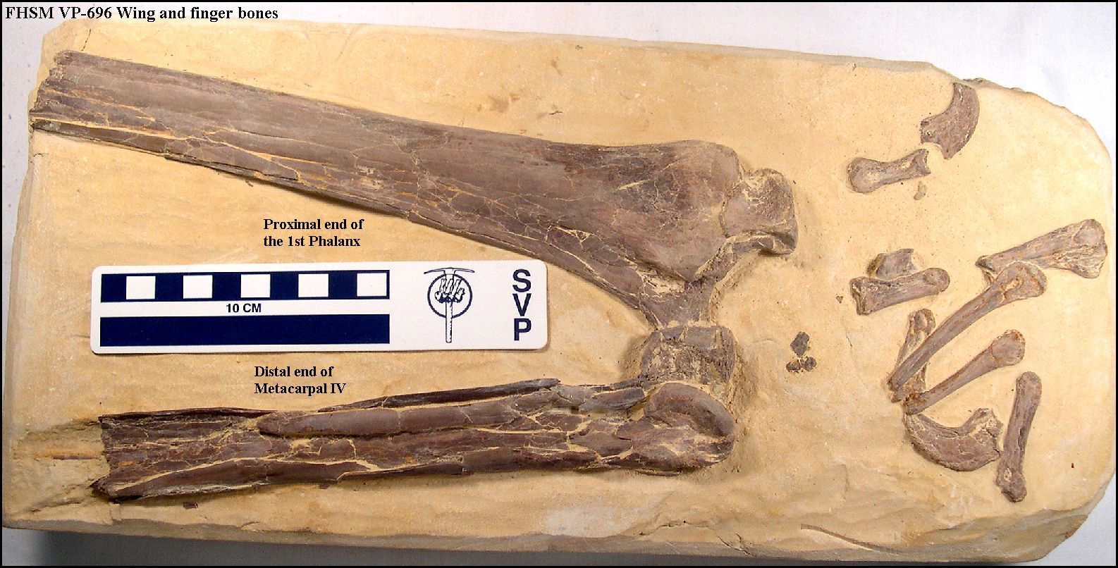 |
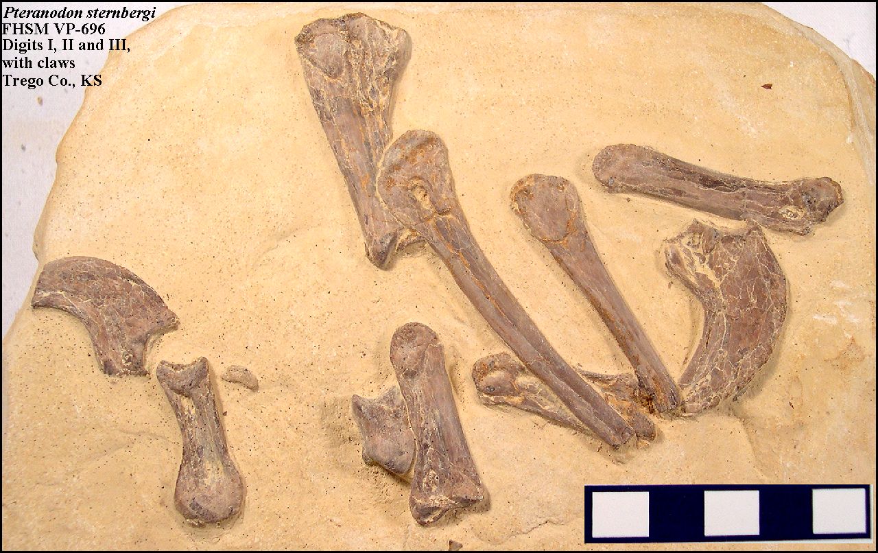
LEFT: A portion of the middle of Pteranodon wing (FHSM
VP-696), showing the joint between wing phalanx 1 (upper) and metacarpal IV (lower), and
most of the bones of the pterosaur's "hand."
RIGHT: A close-up of the bones of digits I, II and III as preserved in FHSM VP-696. Specimen collected in 1956 by G.F. Sternberg from southwestern Trego County. |
 |
 |
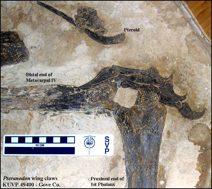
LEFT: Wing fingers (metacarpals) and claw cores (unguals) from a nearly
complete but headless specimen of Pteranodon sp. (FHSM VP-2062) in the Sternberg
Museum of Natural History. The specimen was found by George F. Sternberg in
September, 1956, northwest of WaKeeney, in Trego County. Without the skull, we cannot
determine for certain whether this specimen represents P. sternbergi or P.
longiceps since the post-cranial material is non-diagnostic between these
two species (Bennett, 1992). However, it is most likely P. longiceps since it
occurred in the upper chalk.
RIGHT: Wing fingers and claw cores from KUVP 49400, collected from Gove County by J.D. Stewart. This specimen is figured by Bennett (2001, Fig. 93). |
 |
 |
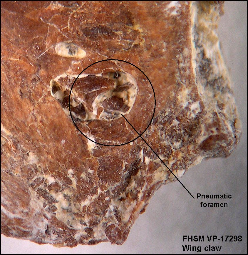
LEFT: Two views of the ungual (terminal digit) for a wing claw
from the right wing of a Pteranodon sternbergi (FHSM VP-17298) that I collected
from southwestern Gove County in 2008. Like much of the rest of the pteranodon's skeleton,
these bone were hollow and filled with air. There is a pneumatic foramen located near the
base of the claw in the picture at far left.
RIGHT: A close-up of the ungual at left showing the pneumatic foramen which would have been connected to the respiratory system of Pteranodon, producing a slight positive pressure within this and other bones of the skeleton. Note that the opening is about 2 mm in diameter. |
 |
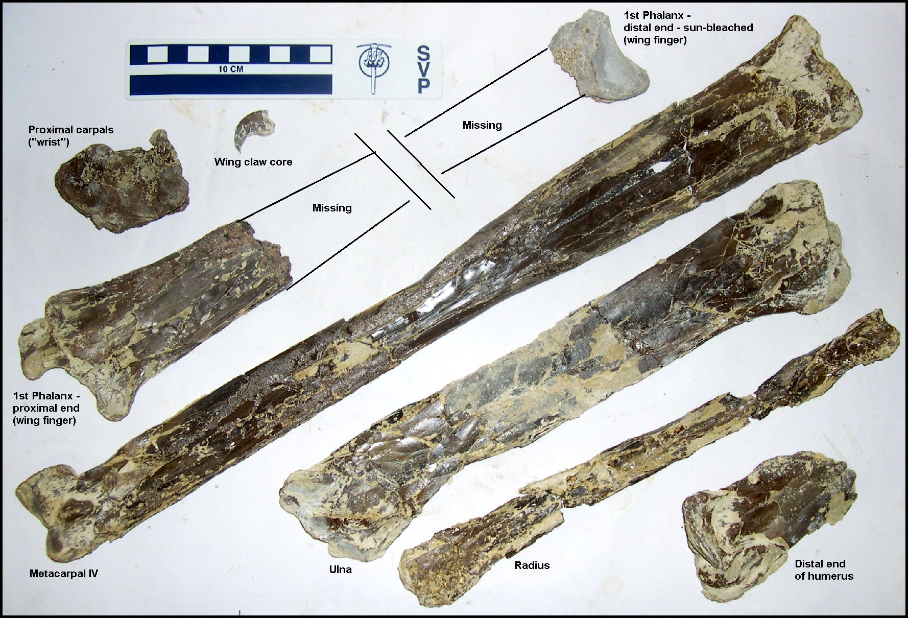 |
LEFT: Wing bones of a Pteranodon sp. that I discovered
in June, 2008 in the lower Smoky Hill Chalk, Gove County, KS. The long bone across
the center of the photo is Metacarpal IV (see drawing above) and is 18 inches (46 cm) in
length. Based on the length of this bone, I estimate that the wing spread of this
pteranodon was about 16 feet. This is somewhat larger than the average female Pteranodon (see Bennett, 1992), so I suspect it was a sub-adult male. Bones recovered so far are shown in yellow.
RIGHT: Proximal and distal views of the left proximal syncarpal of the wing. In Pteranodon, the "wrist" is made up of a pair of fused bones, the proximal and distal syncarpals, which set "back to back" between the radius and ulna, and the large metacarpal IV. The proximal syncarpal articulates with the distal ends of the ulna and radius. The distal syncarpal (not shown) articulates with the proximal end of metacarpal IV (Thanks to Chris Bennett for his help here). |
 |
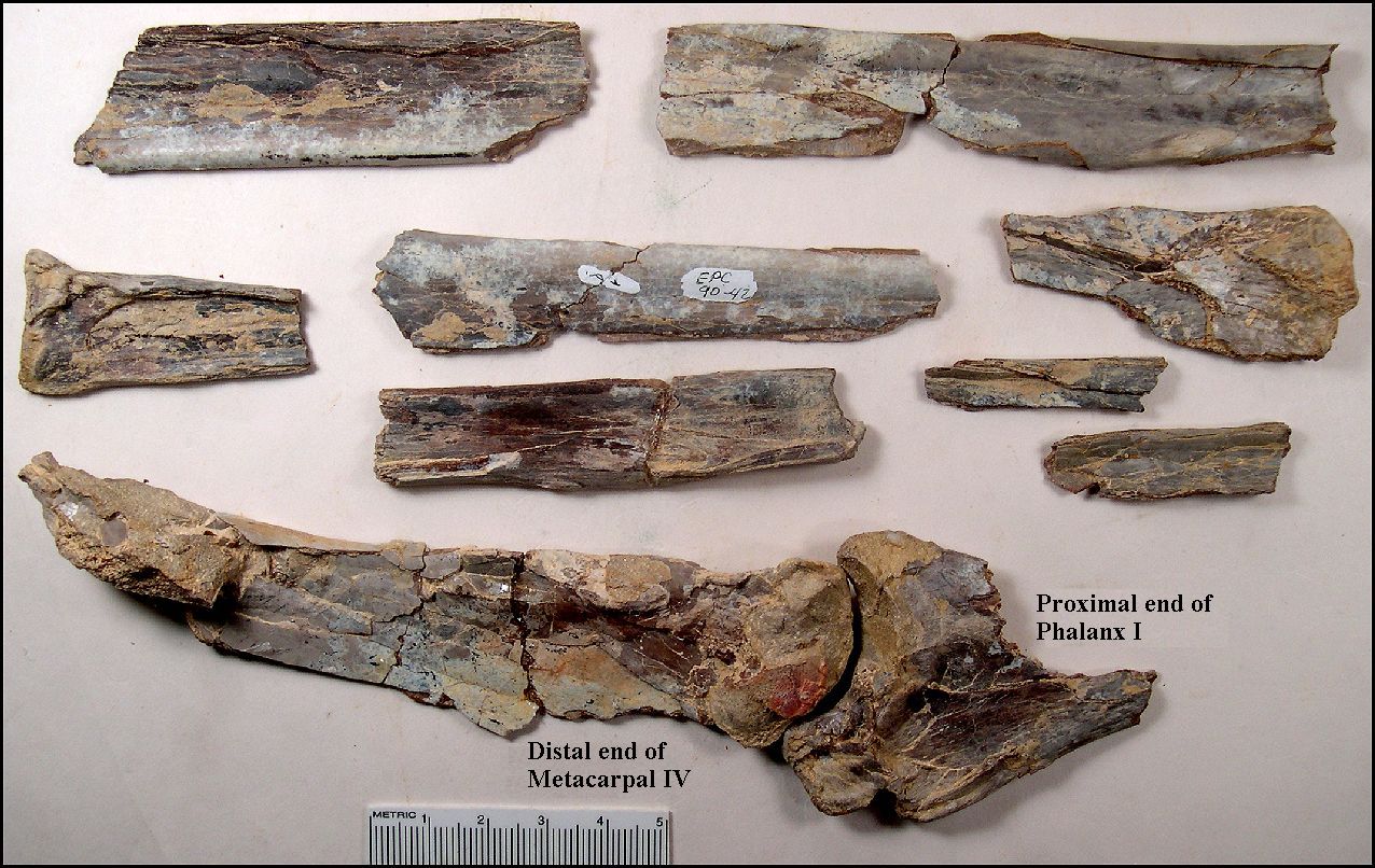 |
LEFT: Most Pteranodon remains that are collected are
fragmentary elements of the wings. EPC1990-42 was surface collected (weathered to a light
blue-gray) and represents the joint between the distal end of Metacarpal IV and the
proximal end of Phalanx I.
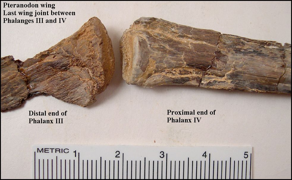
RIGHT: This is an unusual specimen (EPC1990-48) in that represents the end of a wing-finger.... the joint between Phalanx III an Phalanx IV. Click here for a look at the whole specimen. Note that Phalanx IV tapers to a sharp point. |
 |
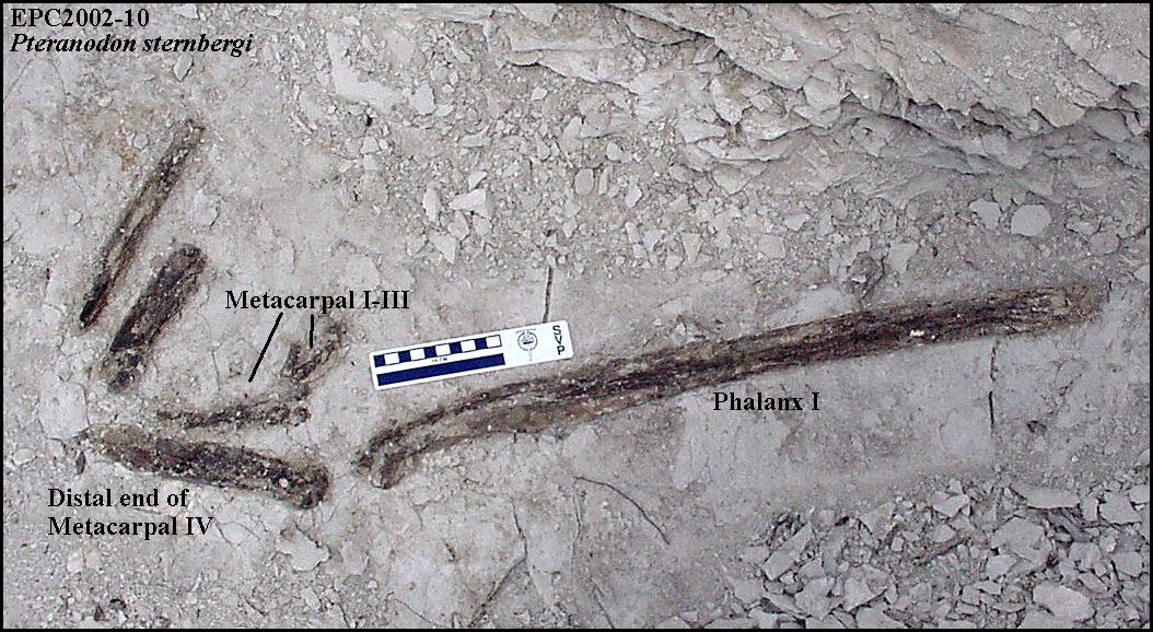 |
LEFT: A field photo of a partial Pteranodon sternbergi wing (EPC2002-10) as exposed in the lower Smoky Hill Chalk of southeastern Gove County.
The specimen shown in the picture, though badly damaged by roots, preserves a complete
phalanx I, a portion of metacarpal IV, and metacarpals I-III, plus some of the
tendons associated with MC I-III.
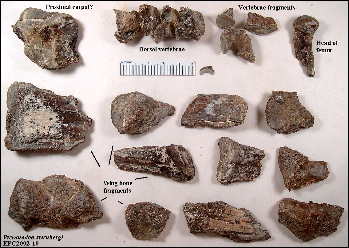
RIGHT: Some of the smaller pieces of EPC2002-10 that were recovered as float. The fragments include one of the carpals, four dorsal vertebrae, the head of one femur and various fragments (joint ends) of wing bones. A single Squalicorax sp. shark tooth was also associated with the specimen. Click here for a close-up view of the dorsal vertebrae. |
 |
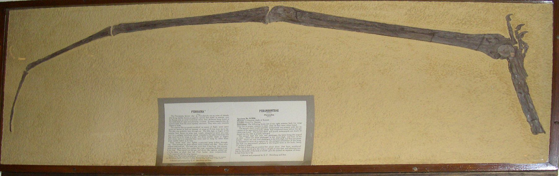 |
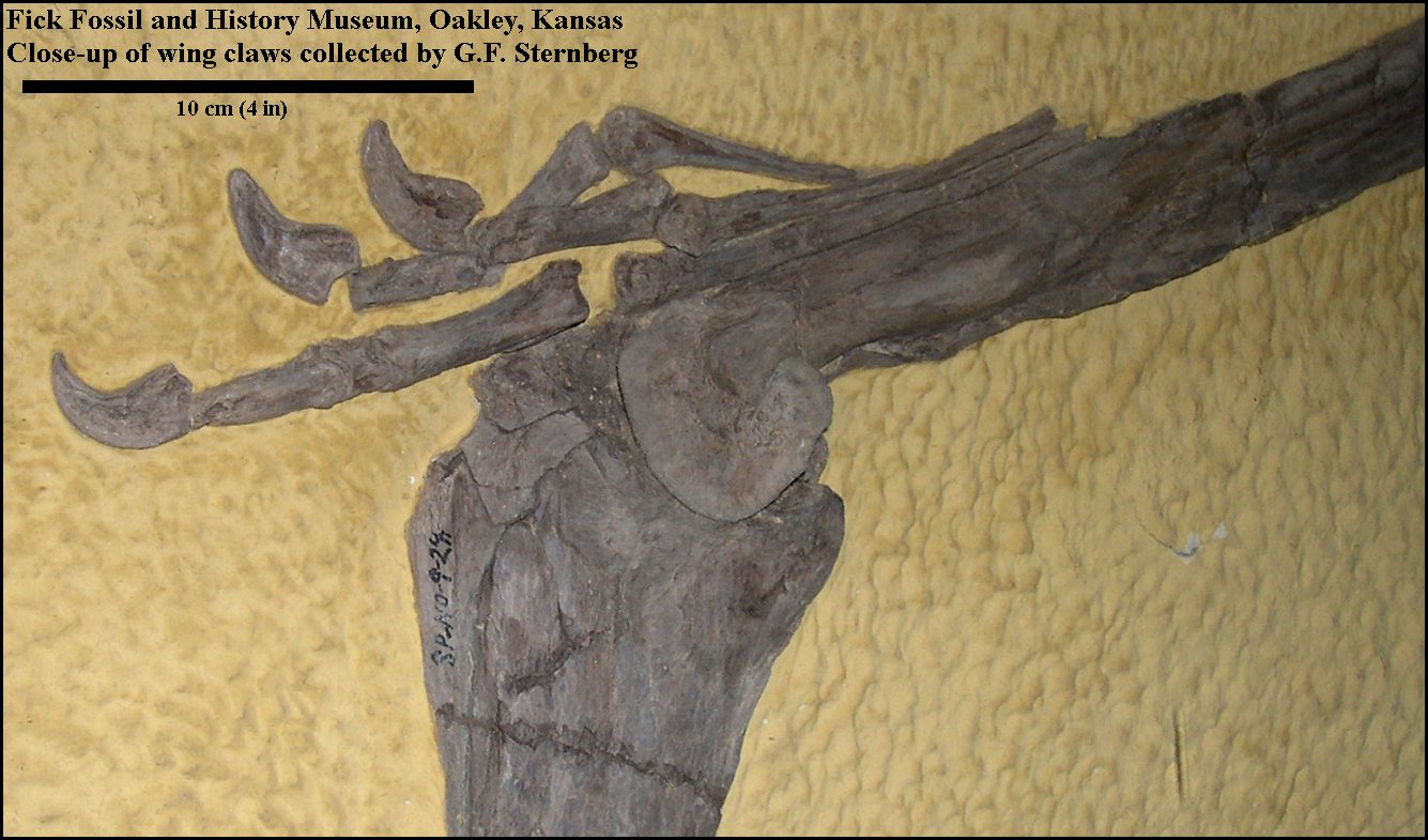
LEFT: A nearly complete left wing (lacking the humerus
and the proximal portions of the ulna and radius) of a Pteranodon (FFHM_1975.438) collected by G.F. Sternberg in September, 1924, and exhibited in the Fick Fossil and History Museum in
Oakley, Kansas. Sternberg's field notes indicate that the recovered portion
of the wing is about 6 ft. long.
RIGHT: Detail of the same wing showing the three digits of the hand and the cores of the wing claws. Note that these bones are crushed. and is essentially two thin layers of bone separated by a thinner layer of soft chalk. Pteranodon bones are not easy to work with. |
 |
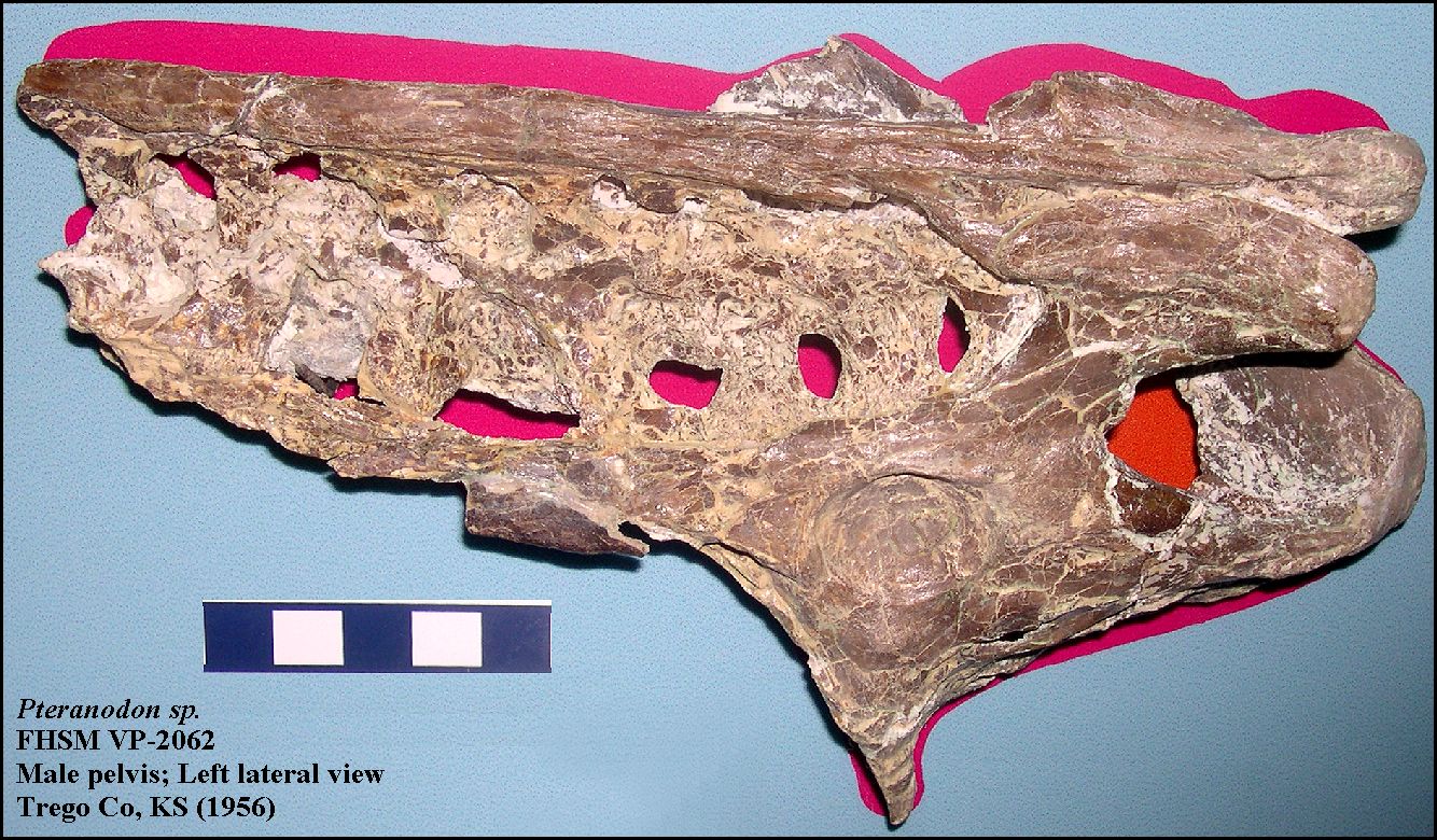 |
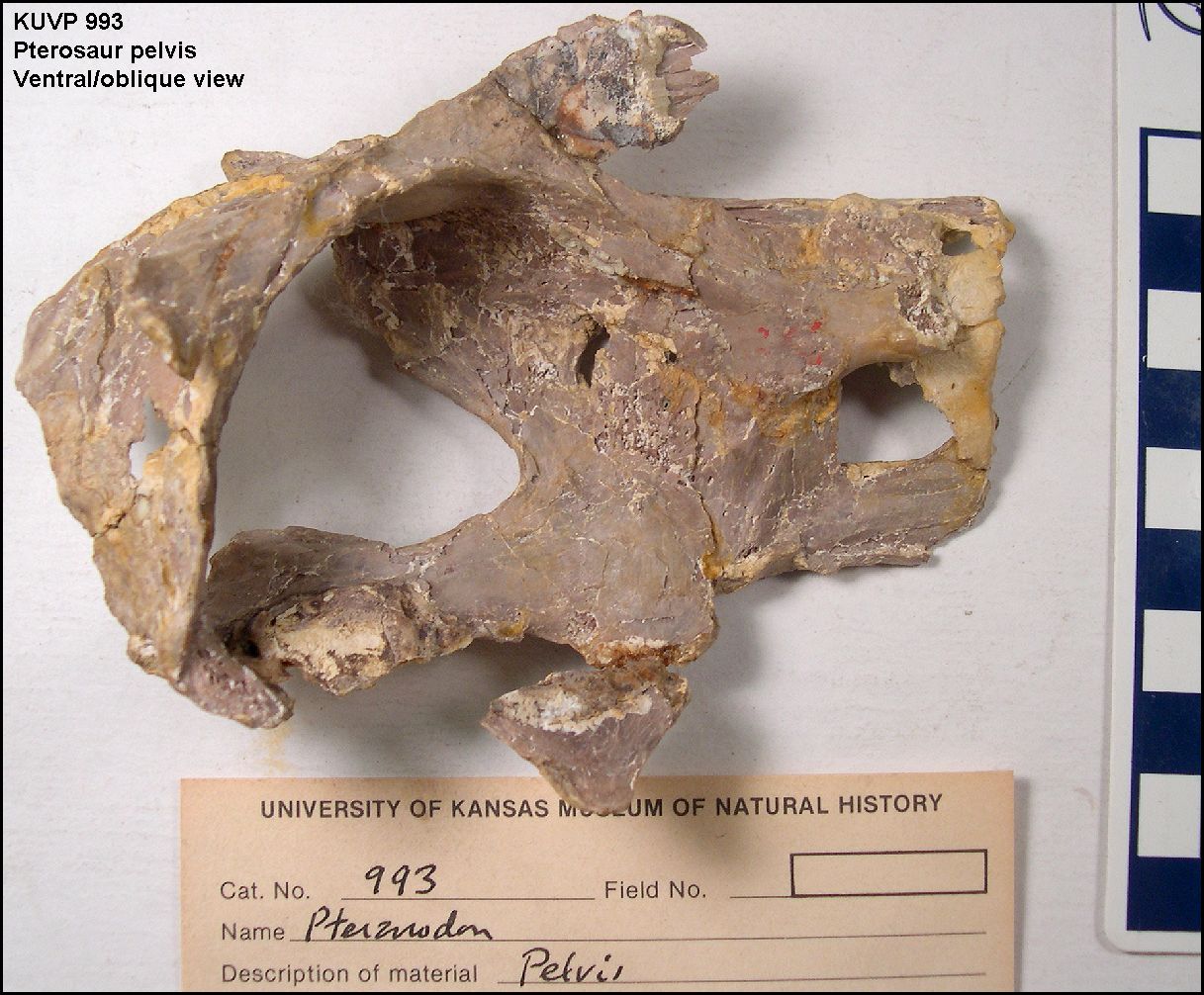
LEFT: The pelvis of a male Pteranodon (FHSM VP-2062) in
left lateral view as shown in the exhibits of the Sternberg Museum of Natural History.
Specimen collected by G.F. Sternberg from Trego County in 1956.
RIGHT: The relatively uncrushed pelvis of a nominal female Pteranodon (KUVP 993; Bennett, 1992, fig.6A) from Gove County in ventral right oblique view showing a relatively large pelvic opening (Anterior to the right). A photo of the same pelvis in dorsal, left oblique view, showing the acetabulum (socket) for the head of the femur.. |
 |
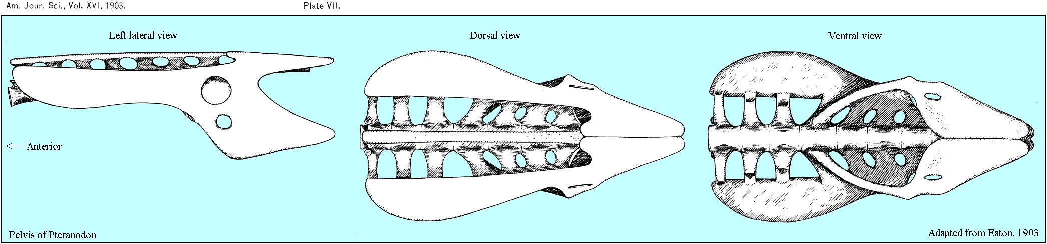 |
LEFT: Three views (left lateral, dorsal and ventral) of the pelvis of Pteranodon, adapted from G.F. Eaton (1903, Plate VII). |
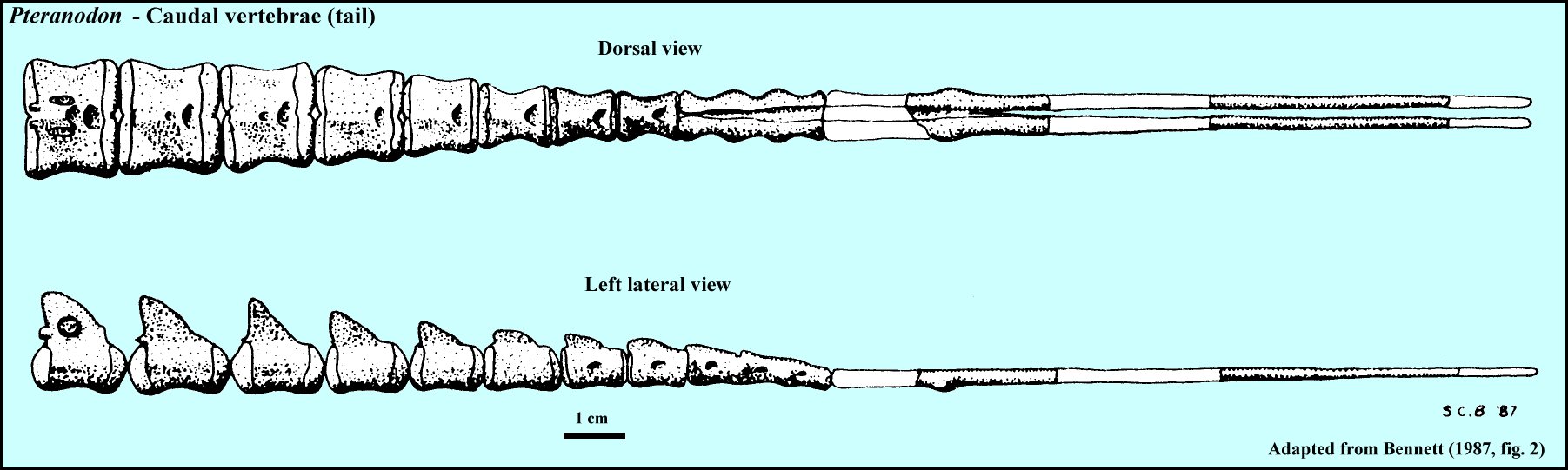 |
LEFT: Pteranodon has a tail (Fig. 2 from Bennett, 1987).... a very short one compared to some Jurassic pterosaurs, but they do have a tail. It is so small that it is rarely preserved. According to Bennett (2001) it is composed of 10 vertebrae, the anteriormost is usually fused to the sacrum. |
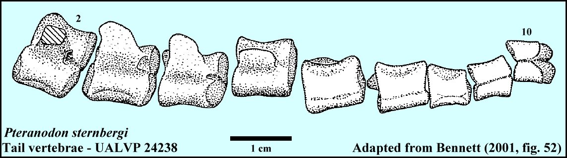 |
LEFT: A drawing of the caudal vertebrae of UALVP 24238 adapted from Bennett (2001, fig. 52). Bennett noted that the last four vertebrae are preserved upside down in this specimen. The posterior 1/2 of the tail consists of a single caudal rod (not shown), possibly composed of several degenerate caudal vertebrae... |
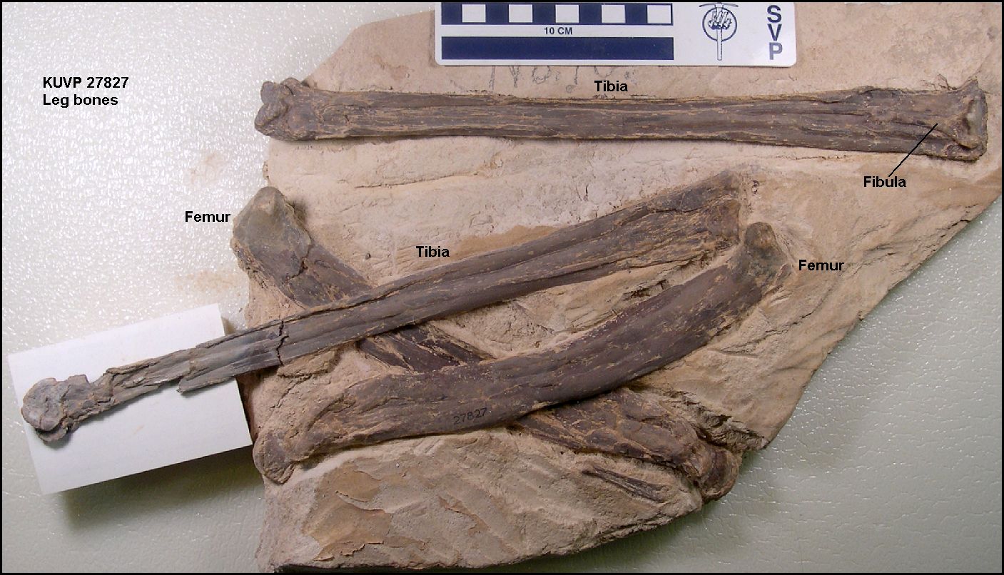 |
LEFT: The paired leg bones (femorae and tibiotarsae) of a Pteranodon (KUVP 27827). Note the splint-like bone (fibula) attached to the tibia at upper right.
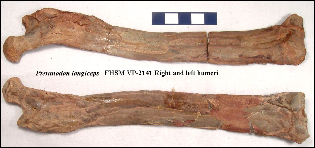
RIGHT: Right and left femurs of Pteranodon longiceps (FHSM VP-2141). Specimen collected in 1950 by R. Taylor near Elkader in Logan County. Length = 23.4 cm. |
 |
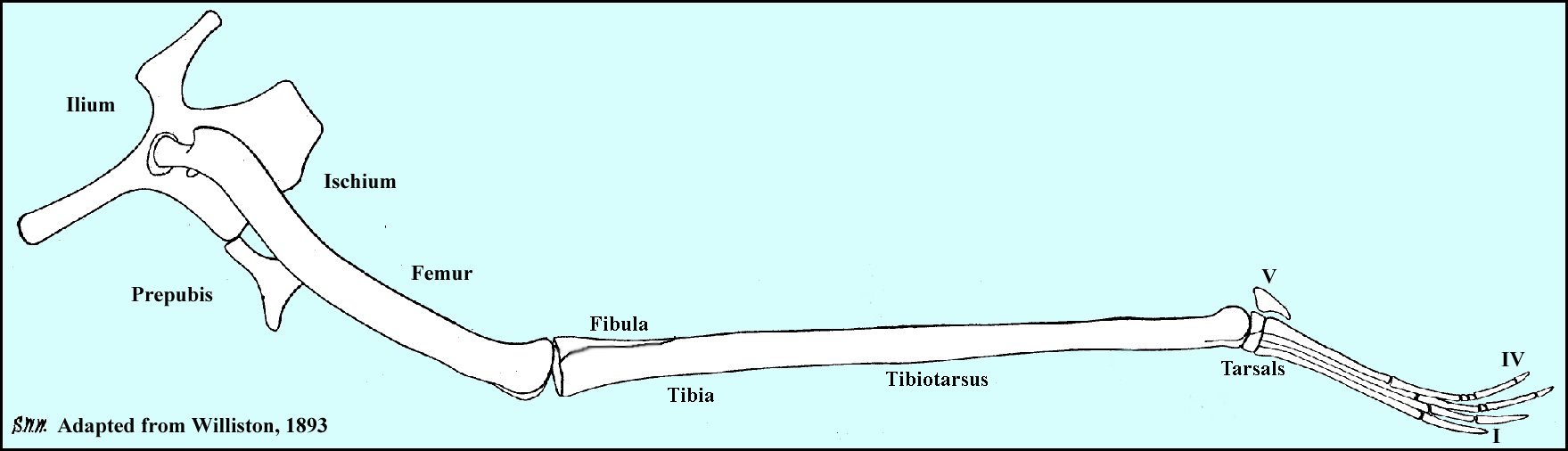 |
LEFT: One of the first drawings of a complete Pteranodon leg and hip (adapted from Williston, 1893). |
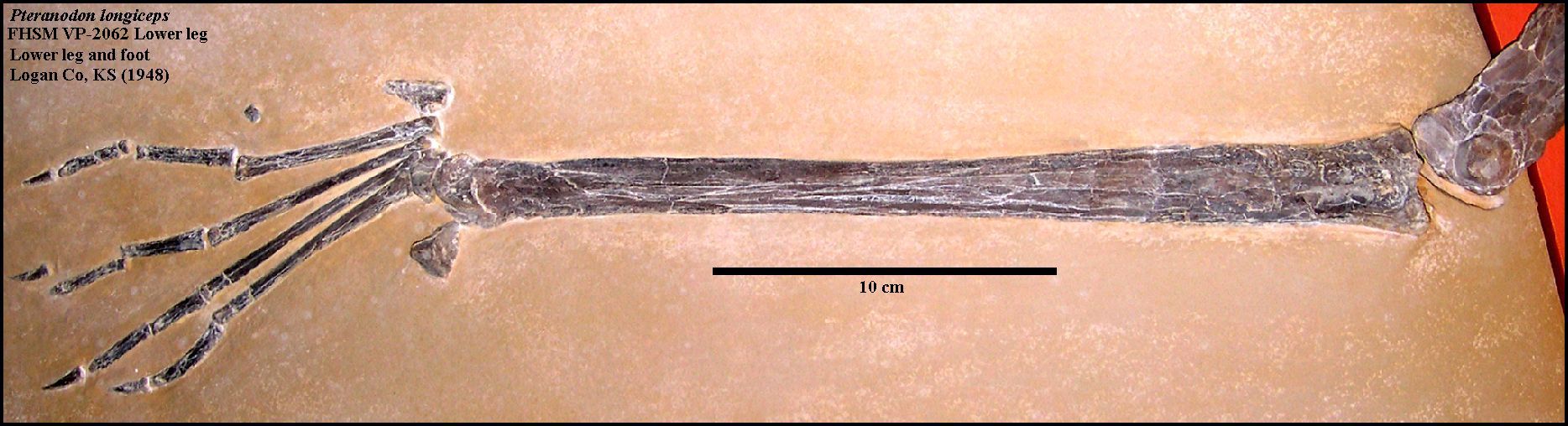 |
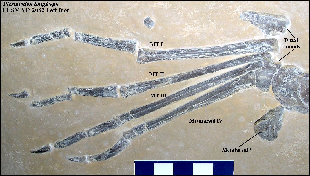
LEFT: Left lower leg (tibiotarsus) and foot of Pteranodon longiceps
(FHSM VP-2062). The tibiotarsus is 27.8 cm in length.
RIGHT: A closer view of the left foot of FHSM VP-2062. |
 |
 |
LEFT: The foot of a recently discovered (2011)
specimen of Pteranodon longiceps from the upper chalk of Gove
County, KS - FHSM VP-17997.
RIGHT: The upper tarsal bones of the foot of KUVP 27827. |
 |
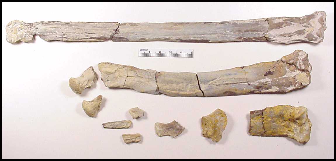 |
LEFT: Photos (2003) of the bones of the right leg of a lower chalk
(MU5) Pteranodon that I collected in 1999 and identified by Chris Bennett. The remains
were discovered by Johan Lindgren. The femur is 22 cm in
length and the tibiotarsus is 30 cm in length.
RIGHT: After some additional preparation and examination, I was able to assemble the complete right femur, and identify the distal end of the left femur, the acetabulum of the hip and a dorsal vertebra. |
 |
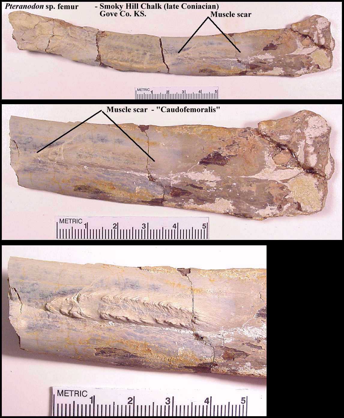 |
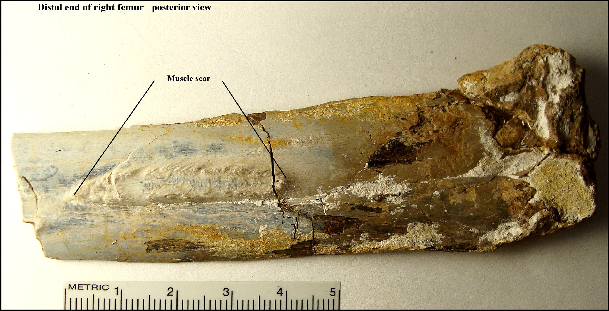
LEFT: The distal end of the right femur shows a very
distinct muscle scar.... most likely the attachment point of the caudofemoralis.
(2003 photo)
RIGHT: Close up (2010 photo) of the same bone, showing the large muscle scar. According to Bennett's 1992 analysis of Pteranodon bones, an average male with a 5.6 m wingspread would has a femur length of 25.0 cm and a tibia length of 32.8 cm while the measurement for a medial female would be 16.4 cm and 24.3 cm respectively. Although there are no characteristics in these bones themselves to distinguish male from female, their size would suggest that these bones were from an older female or a sub-adult male. Pteranodon leg bones are relatively rare as fossils. |
 |
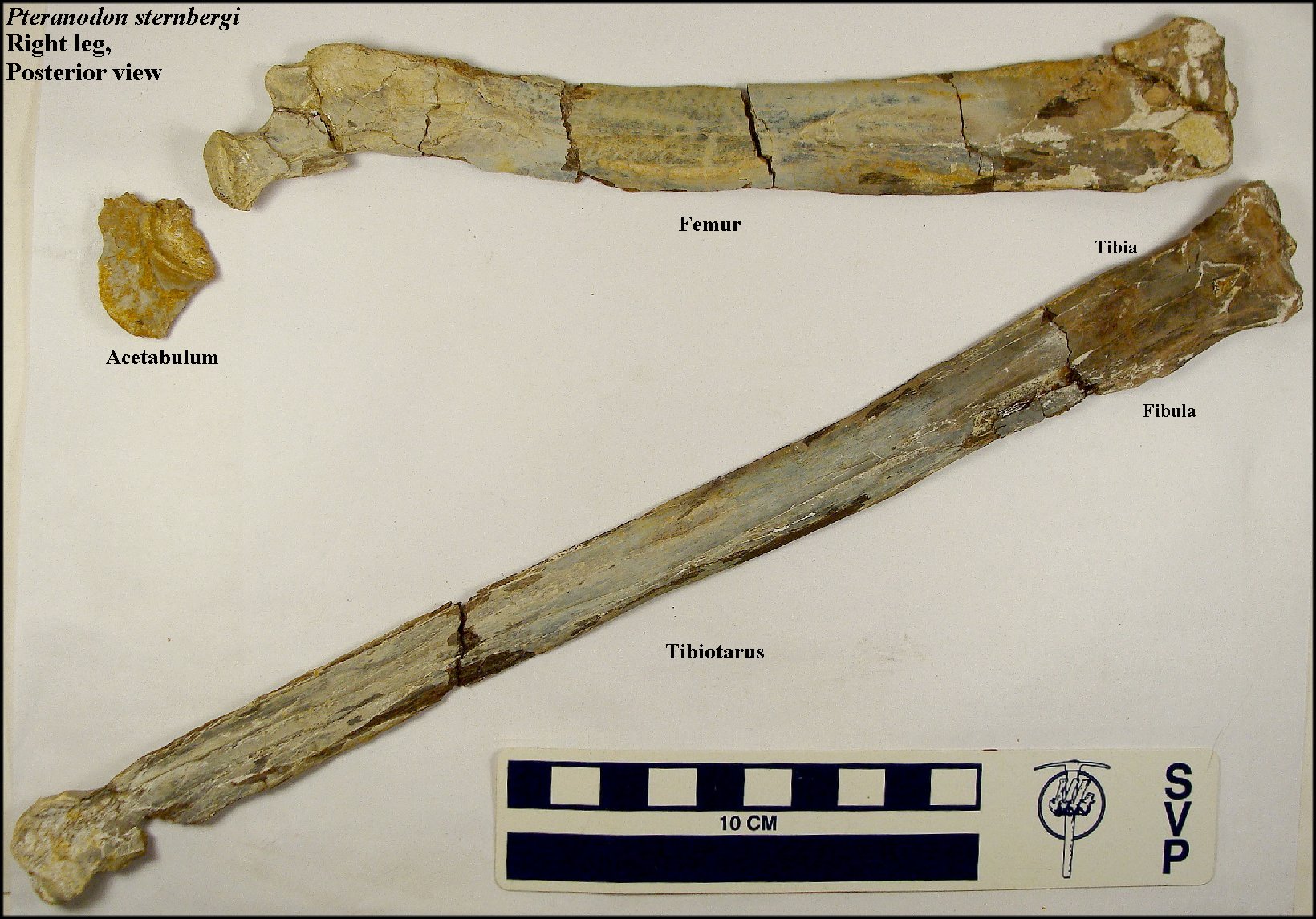 |
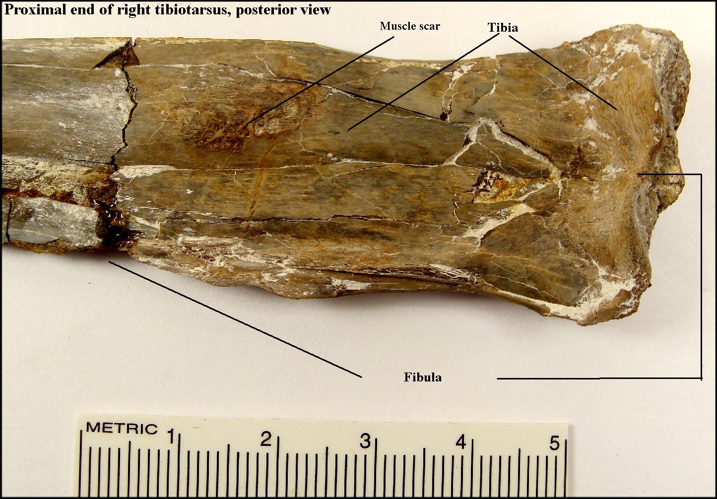
2010 photos:
LEFT: The right leg of Pteranodon sternbergi in posterior view. RIGHT: A close-up of the proximal end of the right tibiotarsus in posterior view showing the location of the fibula. |
 |
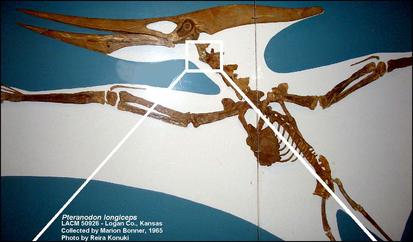 |
UNUSUAL SPECIMEN LEFT: LACM 50926 appears to be a rather normal museum mounting of a large male Pteranodon longiceps in flight. The specimen was collected by Marion Bonner from the upper Smoky Hill Chalk of Logan County in 1965. When you look closely, however, at the top of the neck there is a large Cretoxyrhina mantelli tooth lodged in one of the vertebrae. Whether it was there originally or was added later seems to be lost in time.. but it is certainly an unusual specimen. An injury to this specimen was described in Chris Bennett's (2003) paper on pathologies of large pterosaurs. It also has badly deformed right lower jaw. Photo by Reira Konuki, from his Masters thesis (2008), used with permission. |
|
|
LEFT: A close-up of the 3rd cervical vertebra of LACM 50926
showing an "embedded Cretoxyrhina mantelli tooth" in lingual view.
The slender neck and lightly constructed bones of this male Pteranodon
longiceps would not have put up much resistance to the bone-shearing bite of a shark
with that size teeth. In order to have become embedded in this vertebra, the tooth must
have been almost ready to be shed naturally from the shark's jaw. If it was found there
when collected, it would be the first known instance of Cretoxyrhina feeding on a
Pteranodon.
Photo by Reira Konuki, from his Masters thesis (2008), used with permission. |
Pteranodon sternbergi Harksen
Click HERE to see a Pteranodon sternbergi dig in western Kansas
A large pterosaur, with an adult male wing spread of more than 20 feet which ischaracterized by a large, upward pointing crest. As currently recognized, P. sternbergi is found fairly commonly in the lower Smoky Hill Chalk. The skull of the type specimen was collected by G. F. Sternberg in 1952 along the Solomon River near Bogue in Graham County, KS. Pteranodon sternbergiremains first occur in the lower Smoky Hill Chalk near Hattin's marker unit 5. P. longiceps is a closely related species that occurs higher (Middle Santonian) in the chalk.
Note here that Kellner (2010) considers Pteranodon sternbergito be distinct enough from P. longiceps that it should be in its own genus, and has revived Geosternbergia (see Miller 1978). In that case, the specimen should be referred to Geosternbergia sternbergi. I am doubtful that his suggestion will be accepted. So far as I am aware there are no other Pteranodon specimens known that preserve this unusual crest. This suggests to me that the crest may, in fact, be malformed and the result of a pathology (tumor or injury) during the life of the individual.
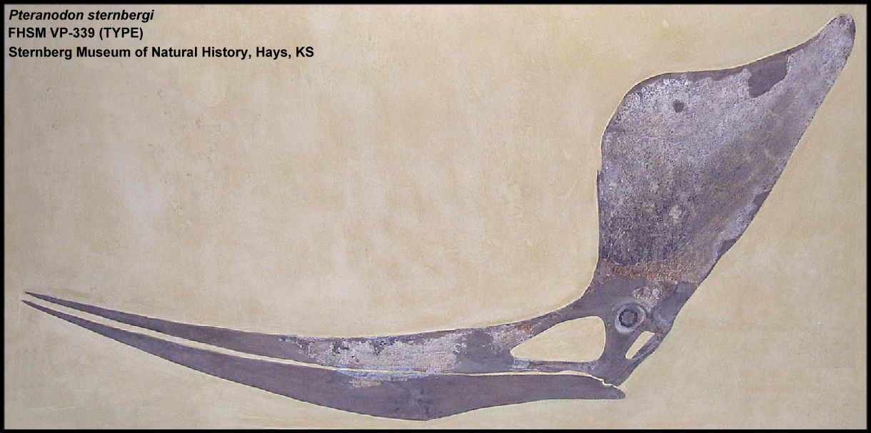 |
LEFT: The partially reconstructed type specimen (FHSM VP-339) of Pteranodon
sternbergi in the Sternberg Museum of Natural History. (Nominal male based on the
size of the skull and the development of the crest). Specimen as reconstructed is about
1.5 m in length. The crest as measured from the back of the lower jaw is about 0.8 m in
height.
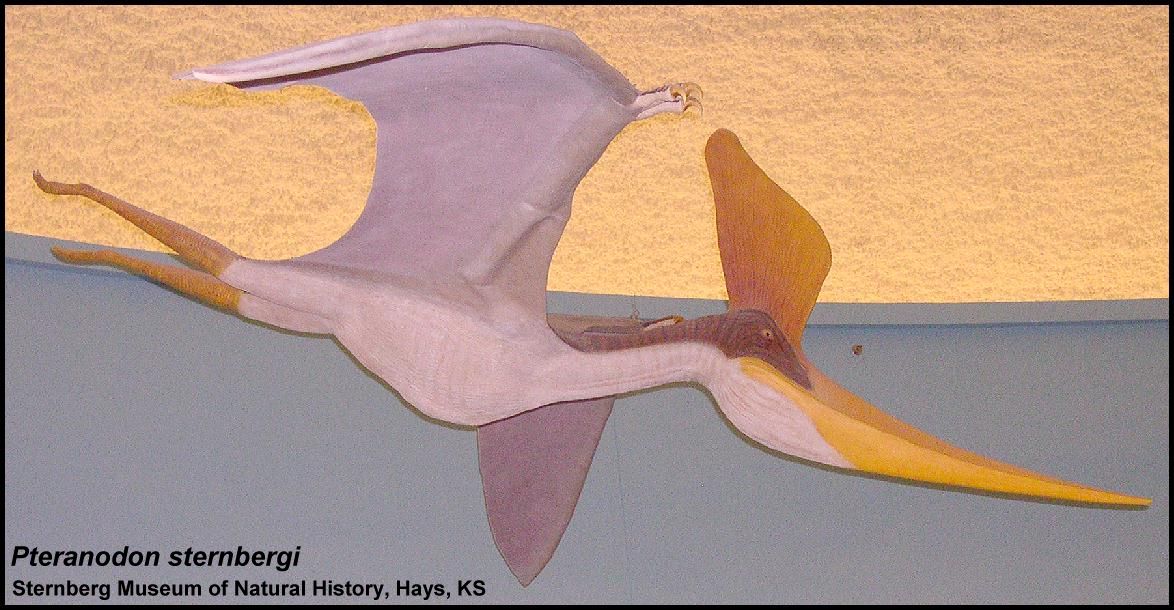
RIGHT: A life sized model of P. sternbergi, also in the Sternberg Museum. |
 |
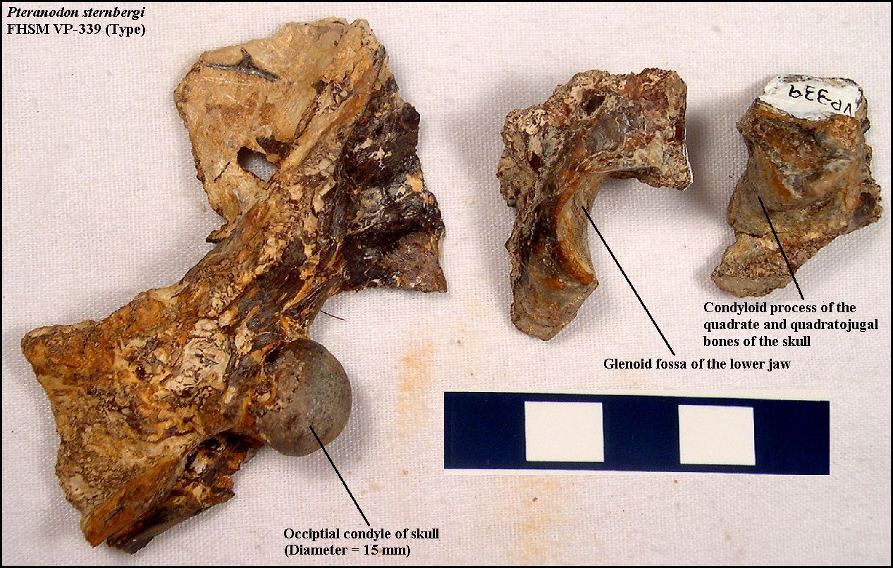 |
LEFT: Fragments of the skull of the type specimen of Pteranodon sternbergi (FHSM VP-339; above). According to G.F. Sternberg, when discovered, the skull had already been damaged by weathering and plant roots. The reconstruction is in part based on the impression of the skull that Sternberg observed when the specimen was collected. The back of the skull, including the occipital condyle, the back of one of the mandibles (glenoid fossa) and the distal end (condyloid process) of the quadrate and quadratojugal were eroded out and collected separately from the larger specimen. |
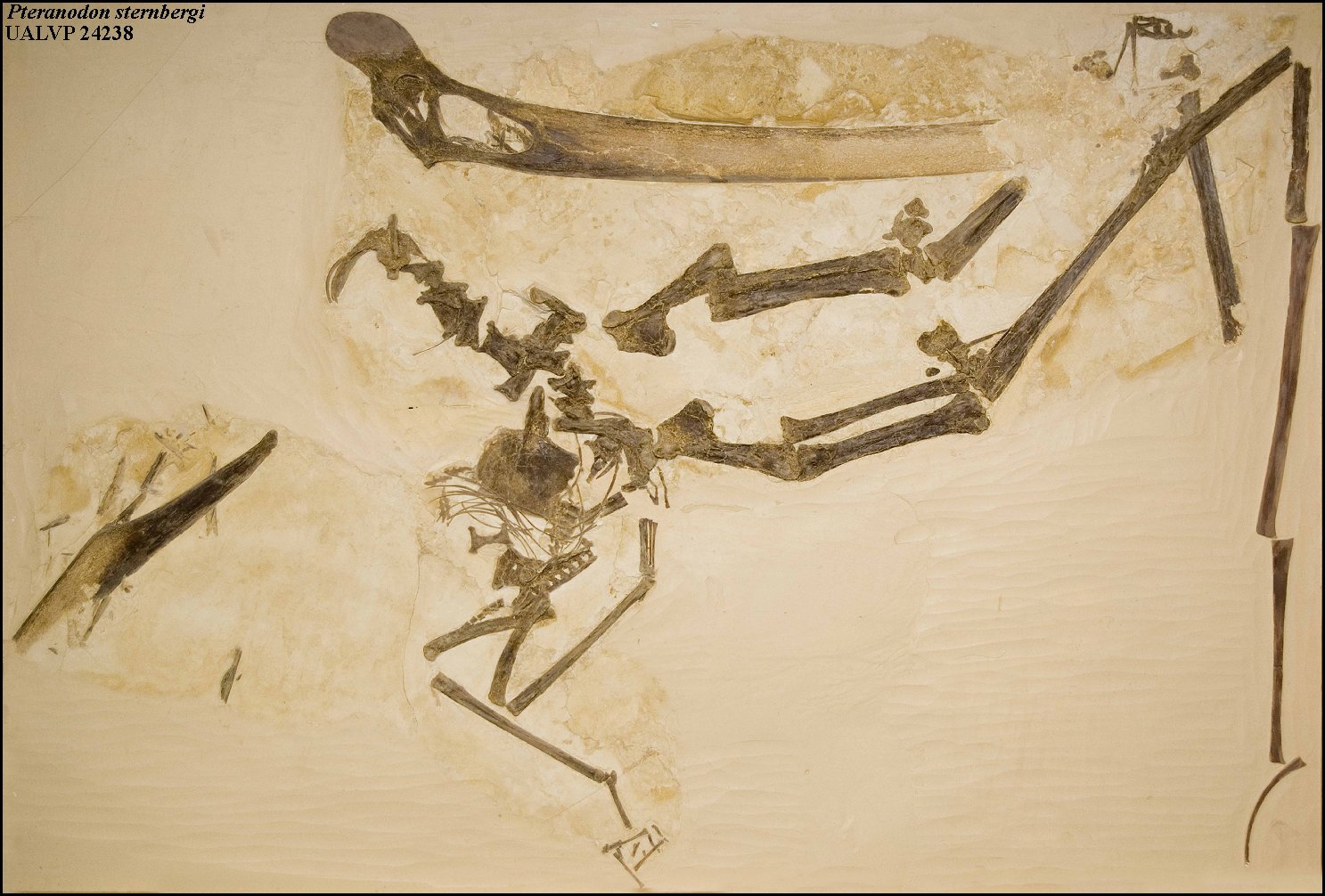 |
LEFT: The Pteranodon sternbergi (UALVP
24238) specimen on exhibit at the University of Alberta was
collected in 1974 from
northern Ness County, Kansas by Dr. Richard Fox and Allen Lindoe of the University of
Alberta, Canada. It is one of the most complete specimens known. Measurements of the wing bones by Bennett (2001) suggest that this
individual was probably a juvenile male with a wing spread of about 4.7 m (15 ft).
Note the Kellner (2010) has designated this specimen as the holotype of a new genus and species - “Dawndraco kanzai” gen. et sp. nov. |
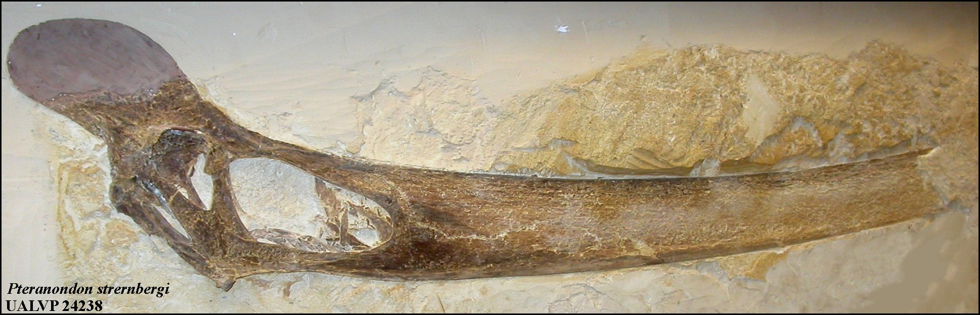 |
LEFT: A detail of the photo above showing the skull of UALVP 24238 which is missing the crest and the anterior portion of the beak. |
 |
LEFT: A drawing of a partial skull of Pteranodon sternbergi (UALVP 24238; adapted from Bennett, 1994, fig. 2) in right lateral view. Although
the skull does not preserve a crest, Bennett described certain features that set it apart
from P. longiceps.
A similarly shaped (appearing to be non-tapering) upper beak (KUVP 967) is in the collection of the University of Kansas and can be viewed HERE. I suspect that the non--tapering appearance is an artifact of being crushed during preservation. |
 |
LEFT: AMNH FR7515 is a nearly complete skull (missing back of the
skull and upper portion of the orbit) collected by Schaeffer and Sorenson from the Smoky
Hill Chalk (MU8-9, middle Santonian) on the Andrew Bird Ranch in Gove County, May, 1952.
Specimen as exhibited is about 76.4 cm (30 in) long. This is a large, male Pteranodon skull. The was the same AMNH expedition that included the discovery of the
"Fish-within-a-fish" specimen by Sorenson.
The specimen is currently on exhibit at the American Museum of Natural History in New York. Photo provided to G.F. Sternberg by Bob Schaeffer in January, 1953. |
 |
LEFT: A recent (2008) photo by Vincent Smith of AMNH FR75165, including the reconstruction of a male crest. Copyright ©2008 by Vincent Smith. Used with permission. |
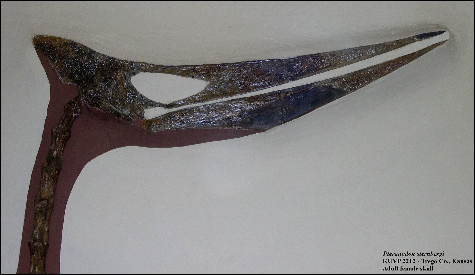 |
LEFT: The nearly complete skull of an adult Pteranodon
sternbergi female (KUVP 2212;
Williston, 1892, Pl. I; see also Bennett,
1992, fig. 3), from Trego County, Kansas. The specimen was collected by
E.C.
Case in 1892, and prepared by H.T. Martin. The skull is 78 cm long. The missing portion of
the lower jaw is colored dark gray. At the time of its discovery, it was the best skull
known.
RIGHT: A portion of the composite Pteranodon specimen on exhibit in the University of Kansas Museum of Natural History. |
 |
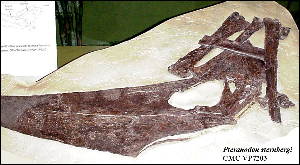 |
LEFT: The nearly complete skull and lower
jaws of a sub-adult male Pteranodon sternbergi (CMC VP 7203) found by my wife,
Pam, in 1996 and later donated to the Cincinnati Museum Center. Note that the end of
the beak was missing when collected, and that the bones around the crest are from one of
the wings that was wrapped around the skull as prior to burial. The radius and ulna are in
front of the skull. The other two bones are probably metacarpal IV and the 1st wing
phalanx. Two small finger bones are lying across the frontal. Note that the crest is still
relatively small on this young male. Based on the size of the wing bones, I estimated that
this individual would have had wing spread of about 5.6 m ( 18 ft). I believe that
this is probably one of the 10 best Pteranodon skulls ever collected from the
Smoky Hill Chalk.
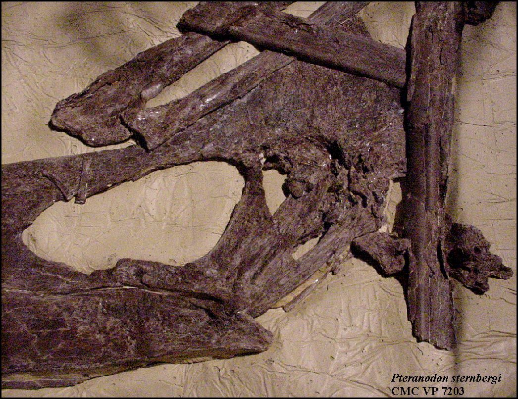
RIGHT: Close up of CMC VP 7203 in left lateral view. showing the large naral fenestra and orbit of the left eye. One or more cervical vertebrae are still articulated with the occipital condyle. Both lower jaws are present and still articulated with the skull. No post-cranial material below the shoulders was collected. |
 |
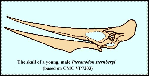 |
LEFT: My reconstruction of what the skull would have looked like
in life. Note that the crest apparently does not become fully enlarged until the male
is fully mature.
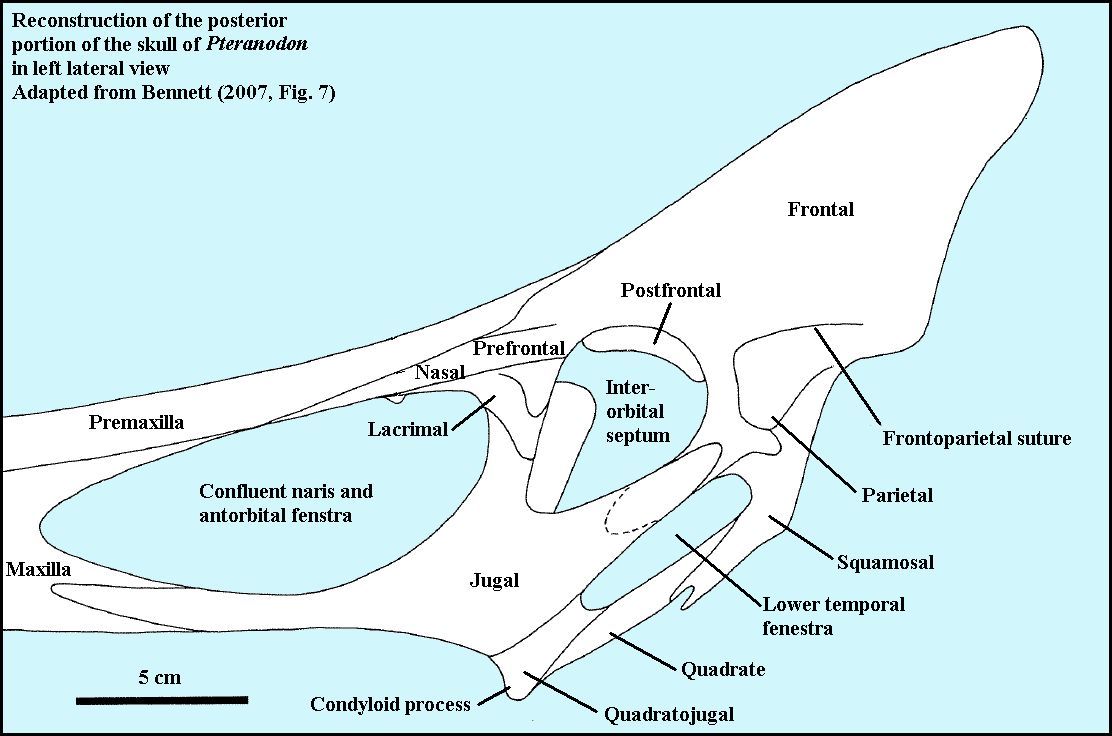
RIGHT: A drawing of the posterior portion of the skull of Pteranodon showing the bones and the openings (fenestra) in the skull. Adapted from Bennett (2001, Fig. 7). |
 |
Nyctosaurus gracilis and N. nanus Marsh
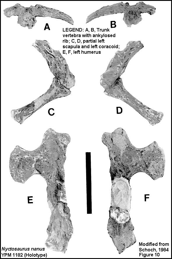
Nyctosaurus (Night Lizard) was a smaller pterosaur that presently occurs only (see note below) in the Smoky Hill Chalk of western Kansas. According the Bennett (1994), there are two valid species; Nyctosaurus nanus in the lower Smoky Hill Chalk and N. gracilis in the upper chalk. N. gracilis was described by O. C. Marsh in 1876 and N. nanus (YPM 1182) was described by Marsh in 1881. Marsh did not figure the specimens but they were shown as photographs by Schoch (1984). Other than size, the skeleton of Nyctosaurusdiffered from P. longiceps most noticeably in the shape of the humerus (upper wing bone), and in the lack of a crest on the skull (Note: see crested Nyctosaurusspecimen below).
Here is the original description of Nyctosaurus gracilis by Marsh (1876): "A second genus of American Pterodactyls is represented in the Yale Museum by several well preserved specimens. This genus is nearly related to Pteranodon, but may be readily distinguished from it by the scapular arch, in which the coracoid is not coossified with the scapula. The latter bone, moreover, has no articulation at its distal end, which is comparatively thin and expanded. The type of this genus is Pteranodon gracilis Marsh, which may now be called Nyctosaurus gracilis. It was a Pterodactyl of medium size, measuring about eight to ten feet between the tips of the expanded wings. Its locality is in the upper Cretaceous of Western Kansas. The type specimens of all the above species are preserved in the Museum of Yale Col1ege."
At the 2000 Society of Vertebrate Paleontology annual meeting in
Mexico City, Chris Bennett mentioned a very large Nyctosaurus specimen:
"A third new specimen from higher in the
Smoky Hill Chalk Member, although incomplete, is the largest known specimen of Nyctosaurus,with an estimated wingspan of 4.5 m. Thus known specimens of
Bennett, S. C. 2000. New information on the skeletons of Nyctosaurus. Journal of Vertebrate Paleontology 20(Supplement to Number 3): 29A. (Abstract)
Note that a new species of nyctosaur (Muzquizopteryx coahuilensis n.gen., n.sp.)was reported from the Austin Chalk of northern Mexico by Stinnesbeck et al. (2005)
 |
LEFT: The holotype specimen of N. gracilis (YPM 1178) was
collected by H. A. Brous on April 21, 1876. It includes the right and left scapulae and coracoids, two
cervical vertebrae, five dorsal vertebrae, another ?dorsal vertebra, ?eighth cervical
vertebra (see Williston, 1903, p. 133), left ulna and radius, right and left carpals,
right and left metacarpal IVs, and right and left first phalanges (Schoch, 1984).
RIGHT: The holotype of N. nanus (YPM 1182) was collected by S.W. Williston on September 22-23, 1876. It includes the left scapula, left coracoid, left humerus, a trunk vertebra with ankylosed rib and other bone fragments (Schoch, 1984). Marsh (1881) originally described the diminutive N. nanus specimen as Pteranodon nanus, but had mistaken the left humerus (E,F) for a femur. |
 |
 |
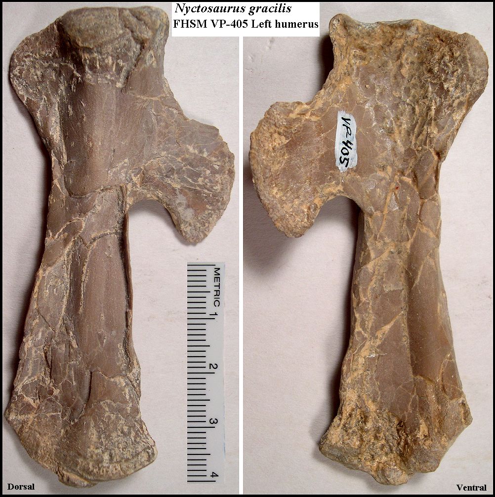
LEFT: Upper wing bones of a Nyctosaurus gracilis specimen (FHSM VP-405) at the Sternberg Museum of Natural History. Note the characteristic
"hatchet" shape of the humerus. Note also that the scapula and coracoid are not fused
in Nyctosaurus in immature specimens like they are in Pteranodon.
VP-405 was collected by Marion and Orville Bonner in 1956 southeast of Russell Springs in
Logan County. It is about the same size as P 25026 (below) in the Field Museum at Chicago.
RIGHT: The left humerus of Nyctosaurus gracilis (FHSM VP-405). Note the distinctive "hatchet" shape. |
 |
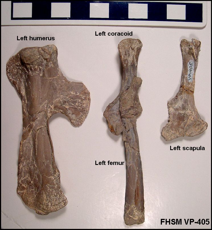 |
LEFT: Limb bones from the left side of the FHSM VP-405 specimen.
Note that the scapula and coracoid are NOT fused in Nyctosaurus like they are in Pteranodon.
In this specimen, the left femur is preserved on top of the left coracoid.
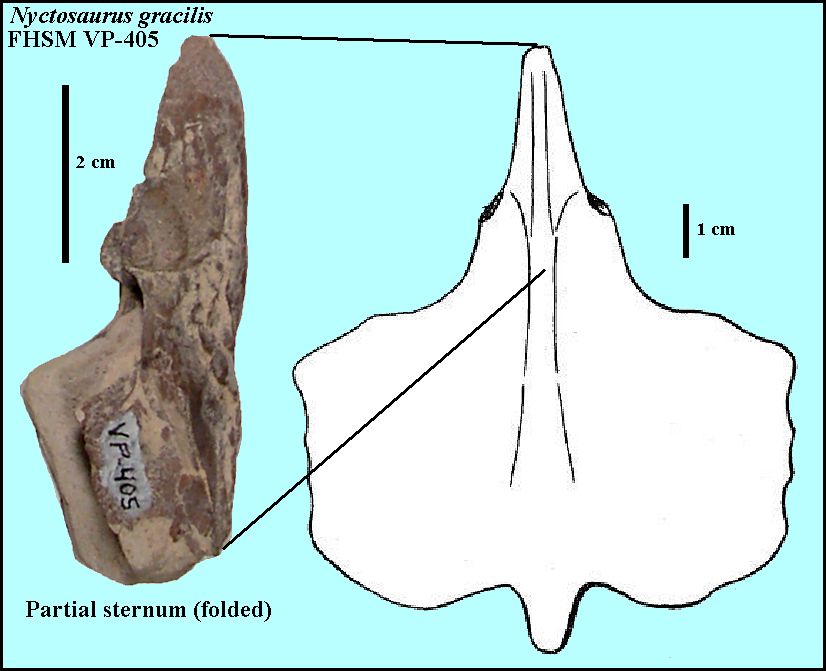
RIGHT: The upper (proximal) portion of the sternum of VP-405. Note that the sternum has been folded along the mid-line in this specimen and is not lying flat as shown in the drawing. |
 |
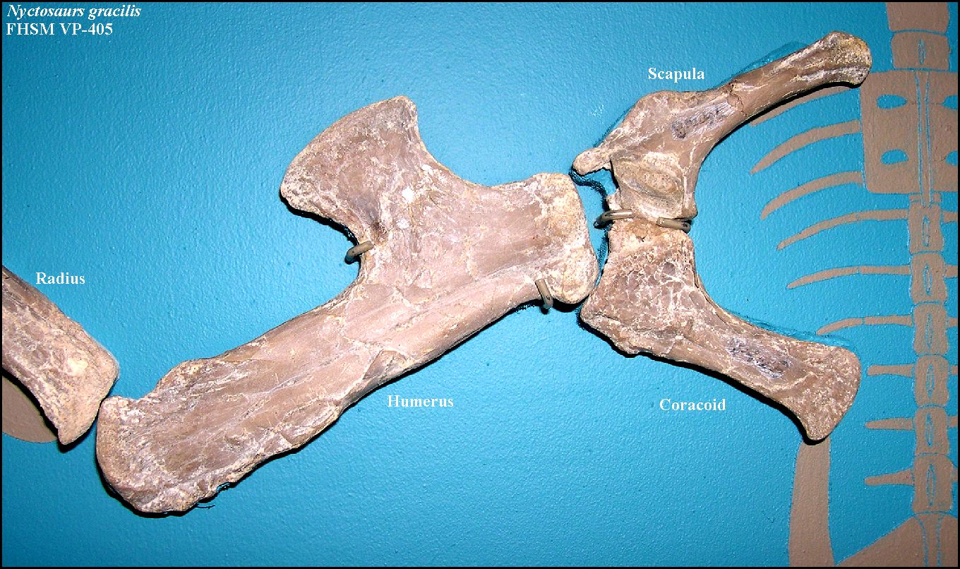 |
LEFT: The right shoulder of FHSM VP-405 as shown on exhibit.
Right: The left humerus of FHSM VP-405. Note the distinctive "hatchet" shape of the humerus in Nyctosaurus. |
 |
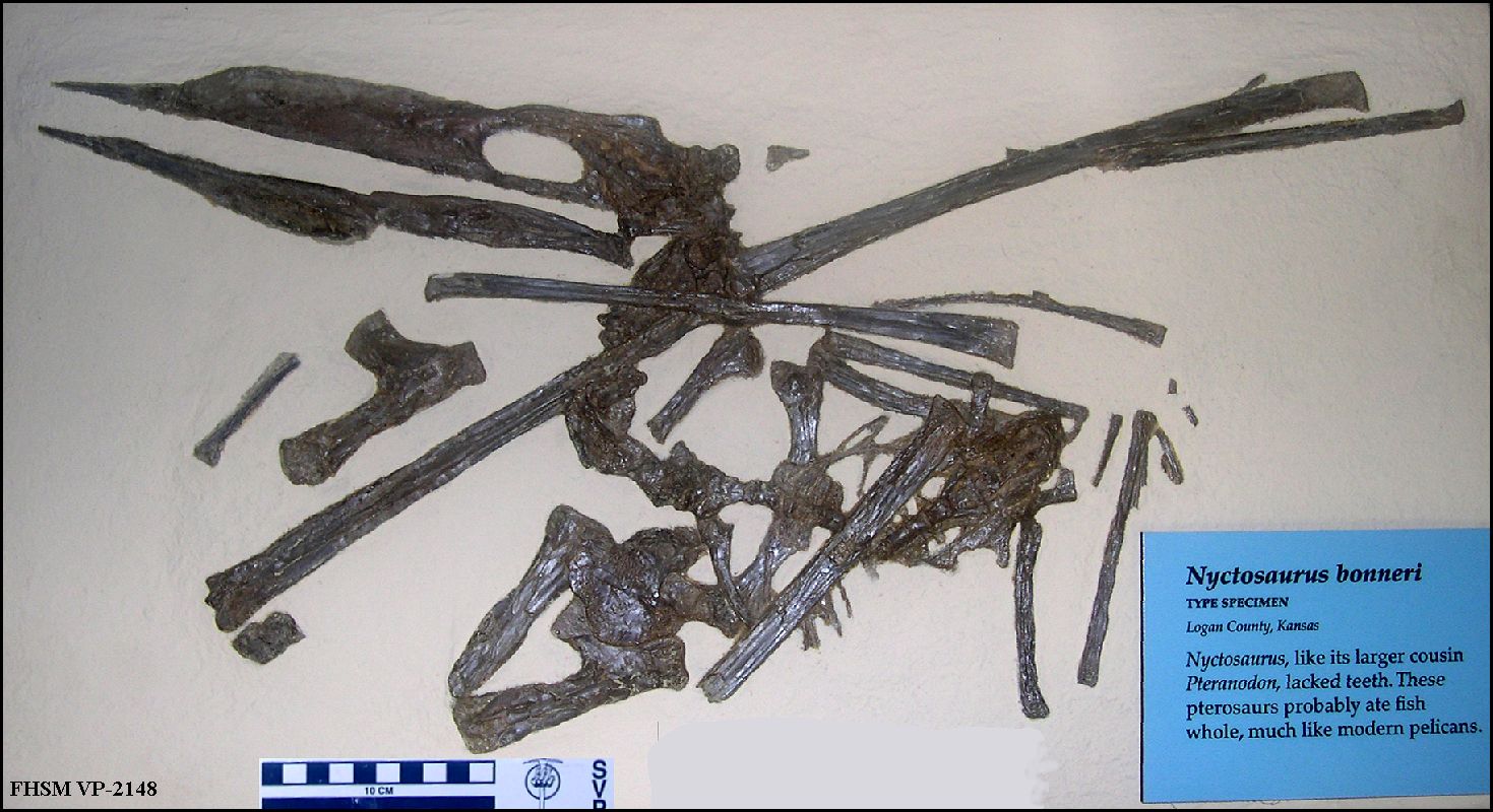 |
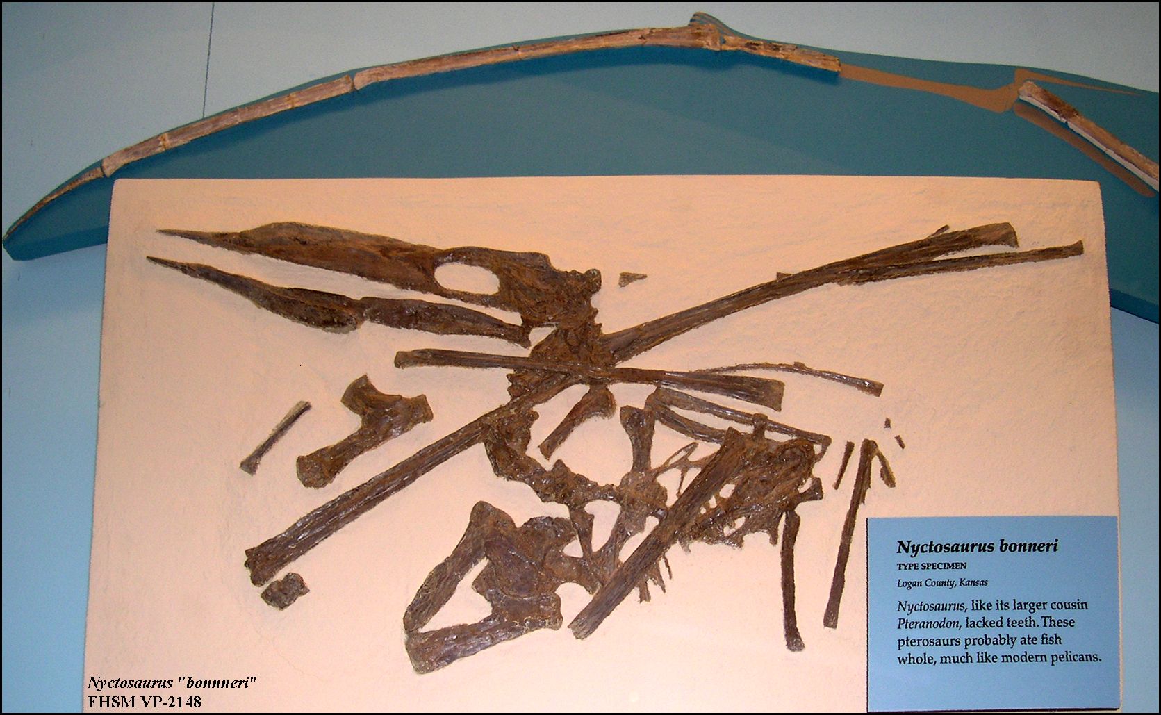
LEFT: The type specimen of Nyctosaurus bonneri (FHSM VP-2148) at the Sternberg Museum, collected from
southeastern Logan County in 1962 by G.F. Sternberg. Bennett (1994) considered N.
bonneri to be a junior synonym of N. gracilis.
Nyctosaurus had 9 cervicals, 12 dorsals, 6 sacrals and at least 3 caudal vertebrae (Bennett 2001). RIGHT: A slightly different view of FHSM VP-2148 showing the distal wing phalanges I, II and III of another Nyctosaurus specimen (FHSM VP-405). Nyctosaurus had one less digit (3) in its wing than did Pteranodon (4). |
 |
 |
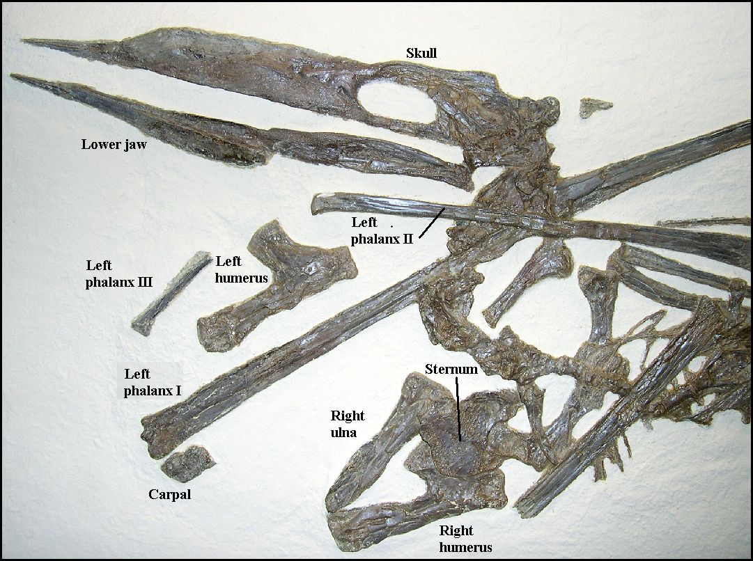
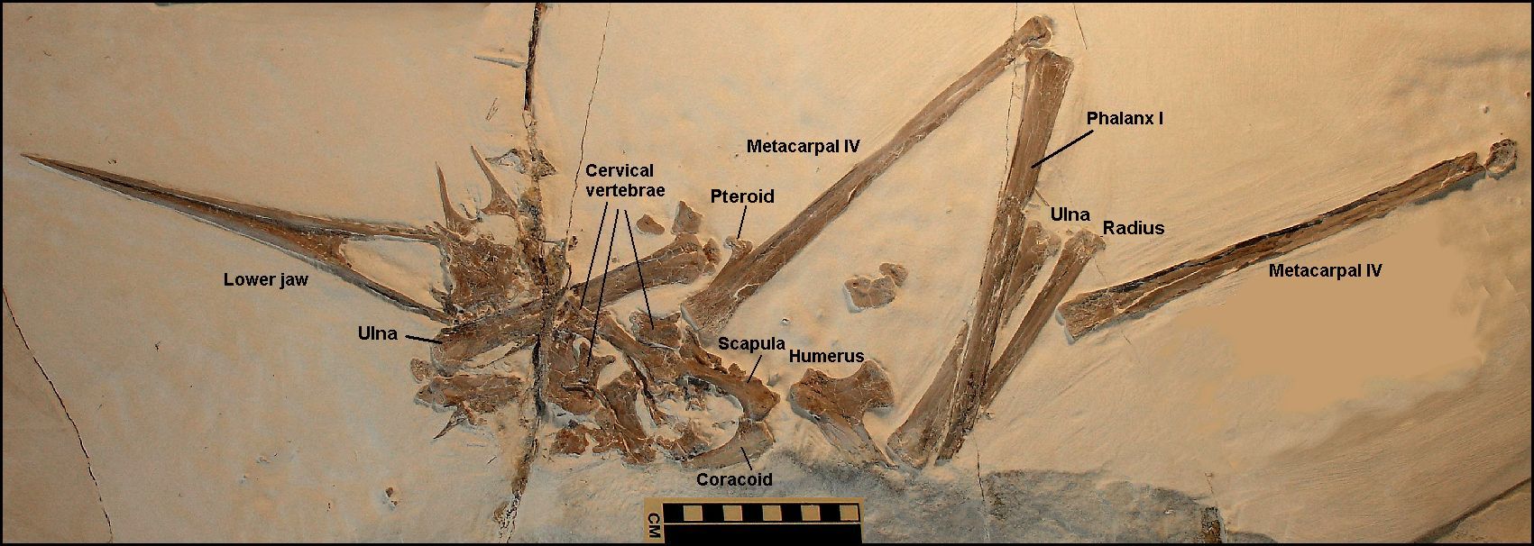
LEFT: The back portion of the skull of . Note that the
large oval opening labeled "naof" is NOT the orbit of the eye... it is the "Confluent naris
and anteorbital fenestra."
RIGHT: The front portion of the skeleton of FHSM VP-2148,with labels. |
 |
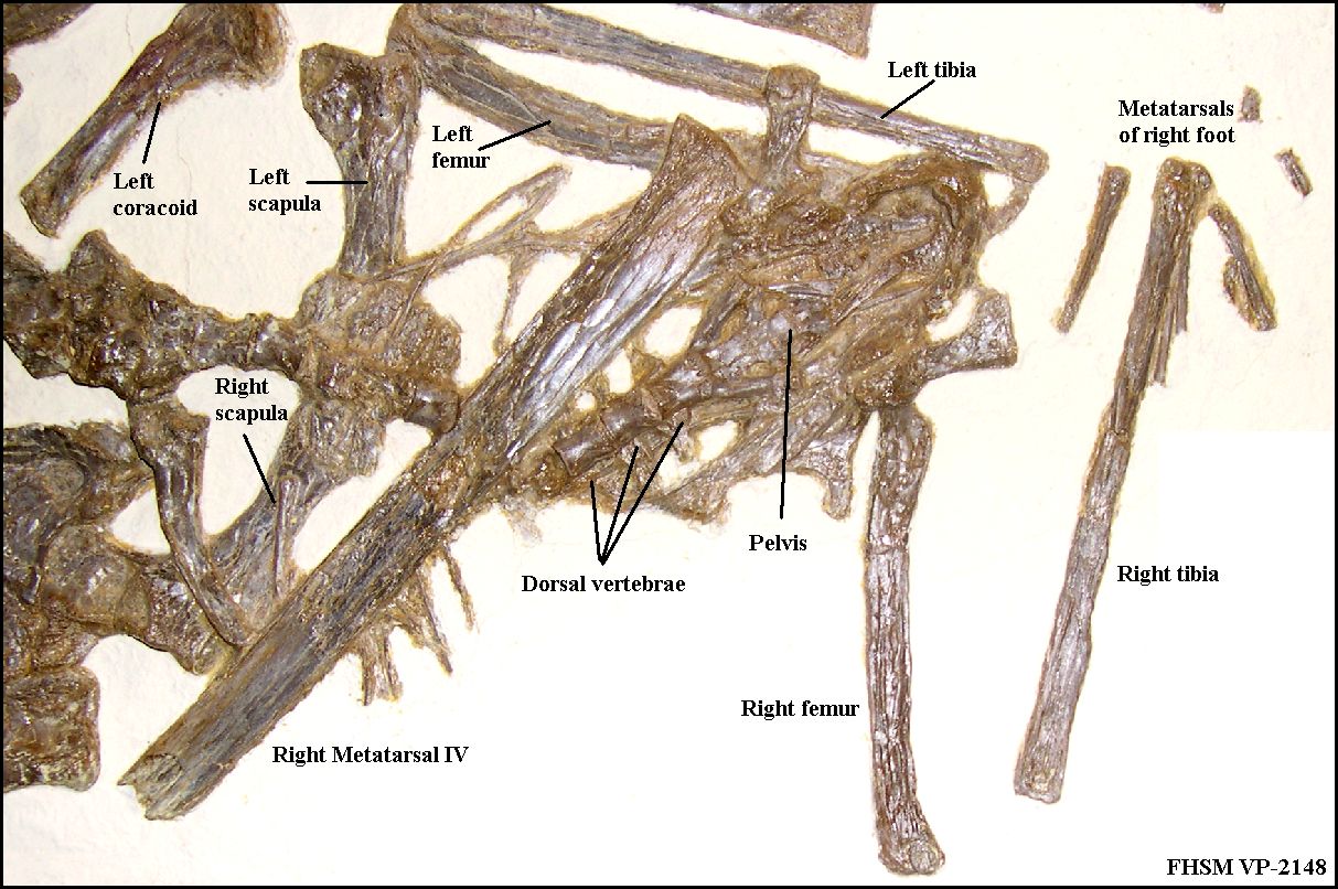 |
LEFT: The posterior portion of FHSM
VP-2148, showing the
pelvis, legs, and dorsal vertebrae. The specimen is shown lying on its back, with the legs
drawn up and the wings folded over the body.
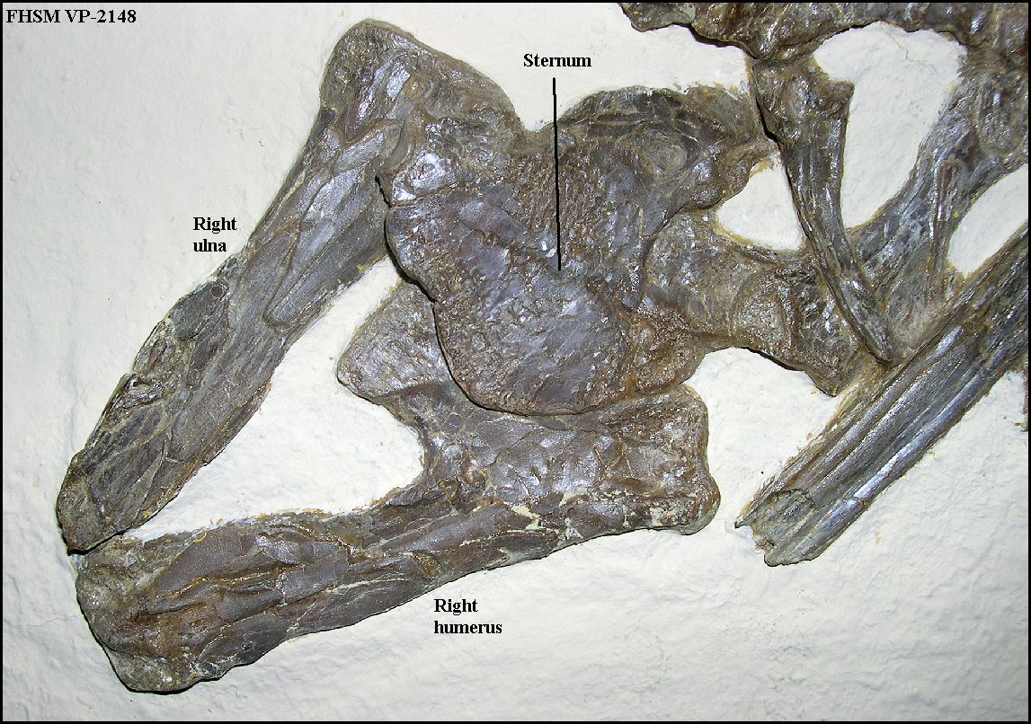
RIGHT: A close up of the center portion of the specimen showing the sternum, right humerus and right ulna. |
 |
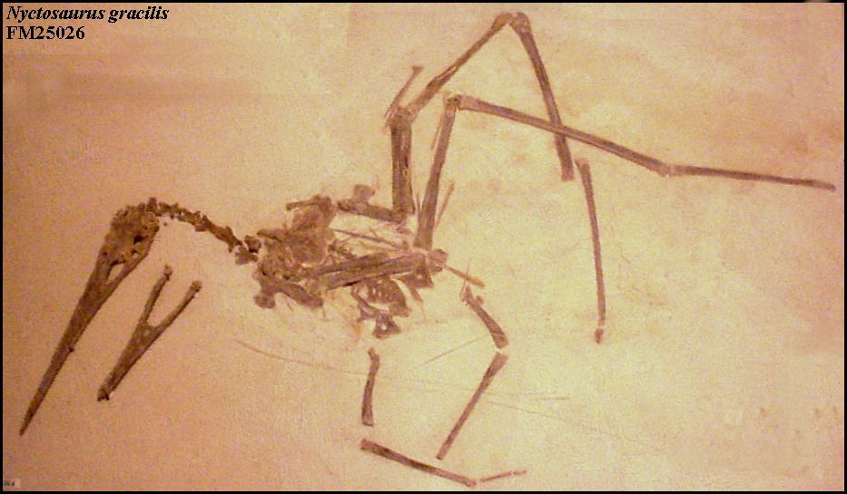 |
LEFT and RIGHT: The first nearly complete specimen of Nyctosaurus gracilis at the Field Museum of Natural History in Chicago, IL. P 25026 was described by Williston (1902). Collected by H. T. Martin in Gove County, Kansas about 1901. (Photographed in 2003) | 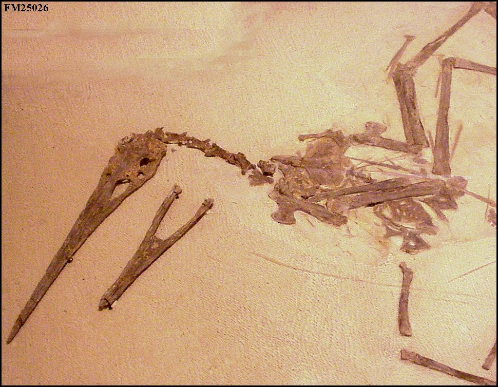 |
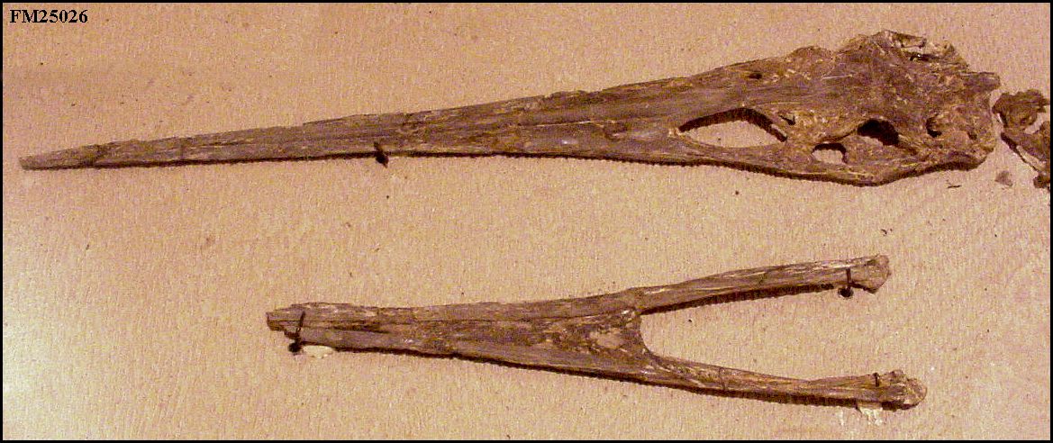 |
LEFT: The skull and lower jaw of Nyctosaurus gracilis (P
25026). This was one of the first pteranodon skulls ever to be photographed for
publication (Williston, 1902).
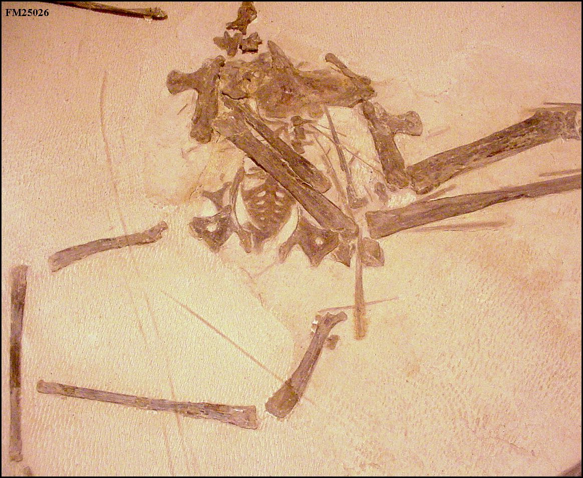
RIGHT: The post-cranial skeleton of P 25026. Click here for a photograph of the lower body of this specimen by Williston (1903, Plate 40) |
 |
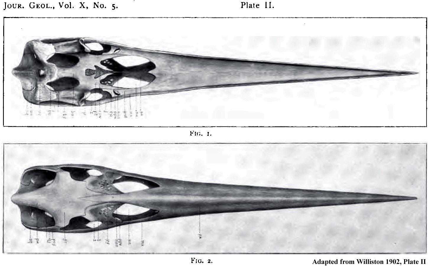 |
LEFT: Skull of P 25026 in ventral and dorsal views,
adapted from Williston (1902, plate II).
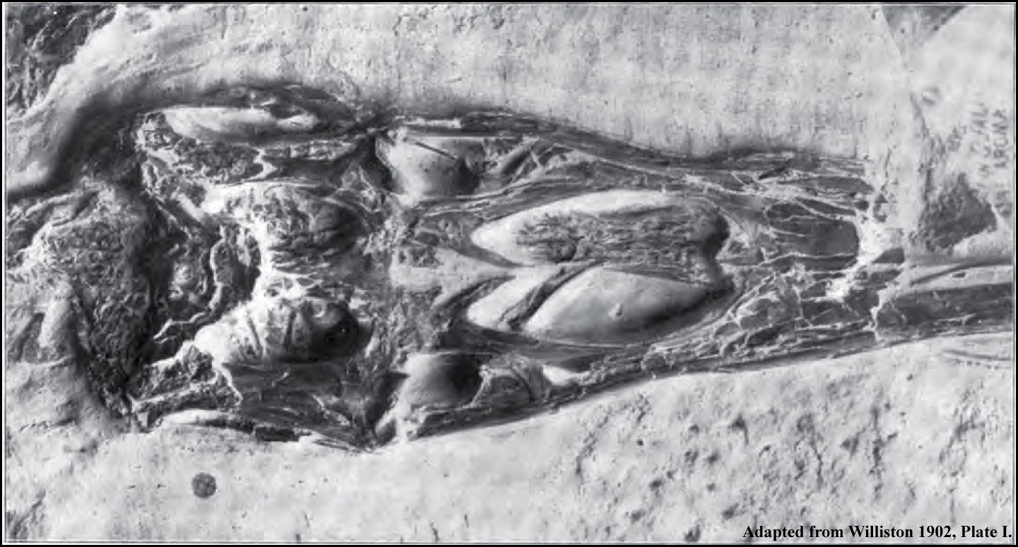
RIGHT: Palate of Nyctosaurus gracilis, P25026, adapted from
Williston (1902, Plate I): Under
surface of the posterior part of skull, lying in matrix slightly enlarged.
Near the anterior end are seen the two hyoid bones, and in the occipital
region, the small triangular pro-atlas. |
 |
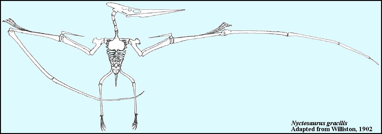 |
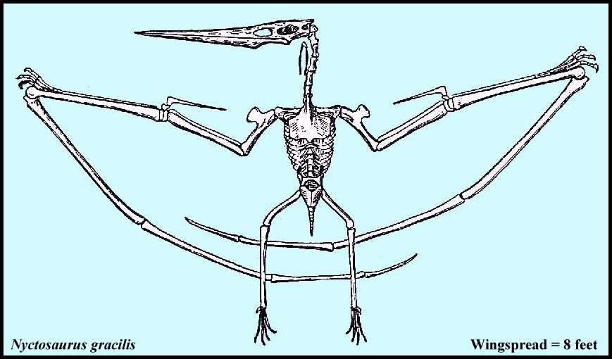
LEFT: The original drawing of the skeleton of Nyctosaurus
gracilis (P 25026) from Williston (1902), based on Pteranodon.
RIGHT: A drawing of the skeleton of Nyctosaurus gracilis from Williston's "Osteology of Reptiles" (1925, p. 299, fig. 190). Until recently, Nyctosaurus was not known to have crests like the larger species of Pteranodon (see below). Also note that Nyctosaurus on only has three wing phalanges, not four as shown by Williston (Brown, 1986). |
 |
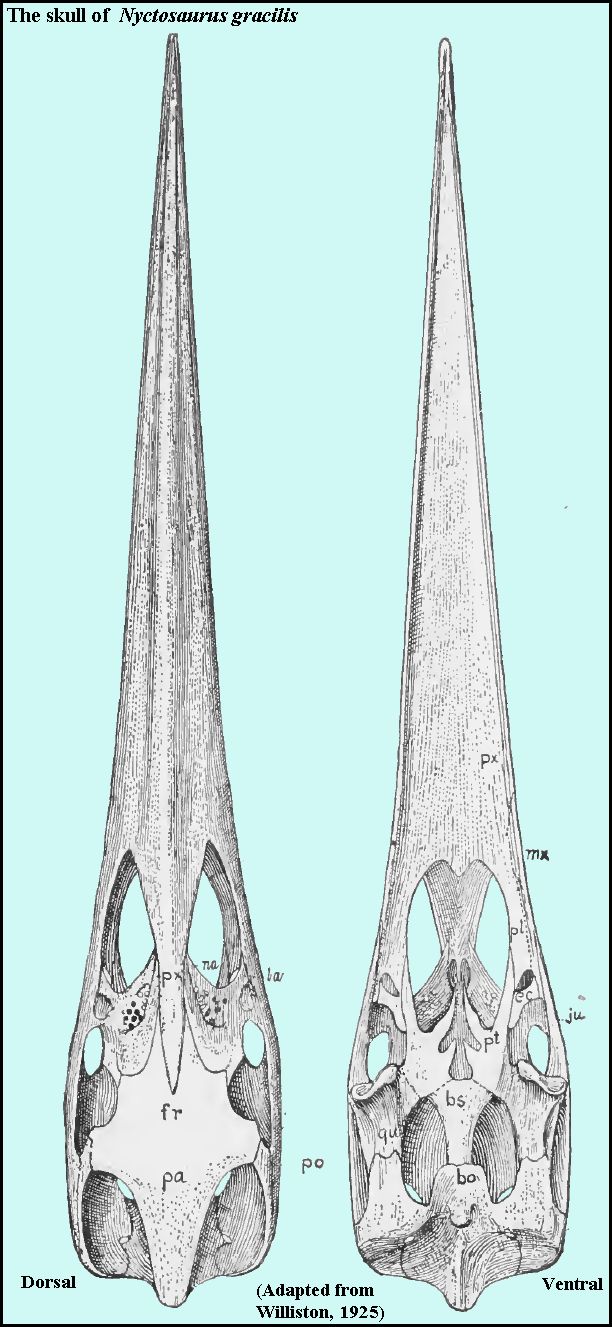 |
LEFT: Dorsal and ventral views of the skull of Nyctosaurus
gracilis Marsh. Adapted from Williston (1925, Pl. 72).
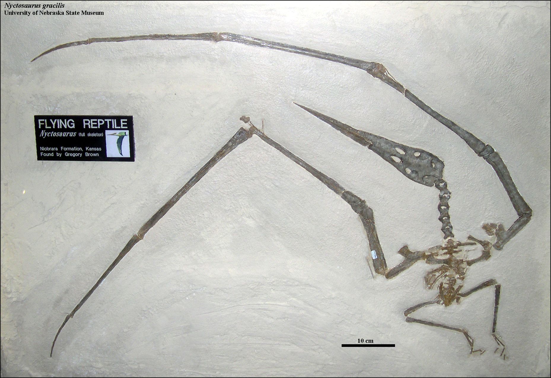
RIGHT: Dorsal view of a nearly complete specimen of a Nyctosaurus gracilis (UNSM 93000) from the Smoky Hill Chalk of Logan County in western Kansas and currently on exhibit in the University of Nebraska State Museum, Lincoln Nebraska. Collected near Elkader, Kansas and prepared by Greg Brown (Brown, 1978, 1986), this specimen is significant because it shows that Nyctosaurus had three wing phalanges, not four as shown by Williston (above). The wingspread on this specimen would have been about 2.4 m (8 ft). Note that what appears to be a wing claw on the left wing is actually a bone fragment from phalanx I. |
 |
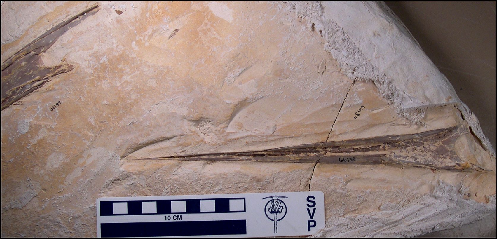 |
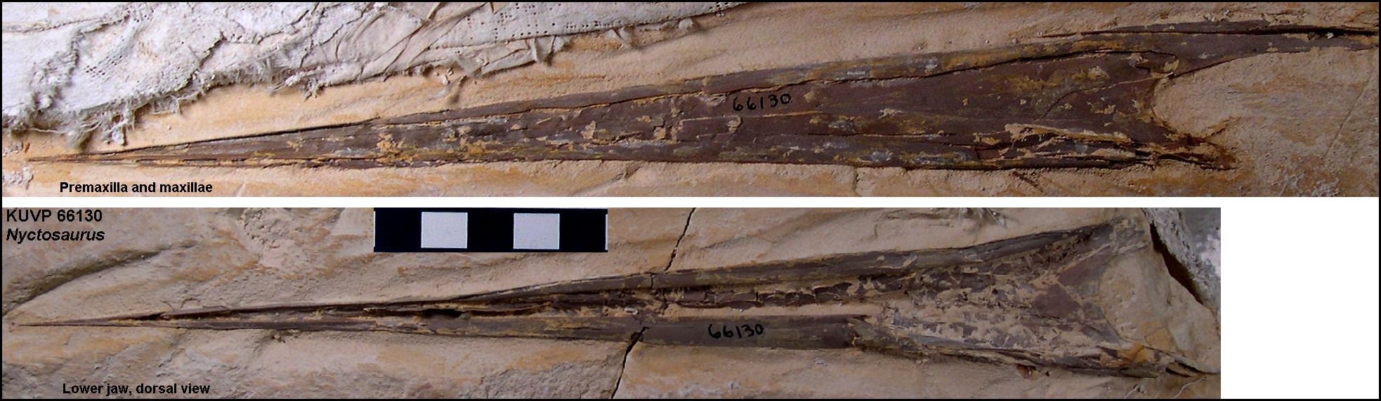
LEFT: A chalk slab containing the upper and lower jaws of a Nyctosaurus (KUVP 66130) collected from the middle Smoky Hill Chalk of western Gove County in 1979.
RIGHT: Photos of both jaws of KUVP 66130 laid side by side for comparison. Cited in Bennett, 2001, p. 33, as a comparison to the Pteranodon dentary.) |
 |
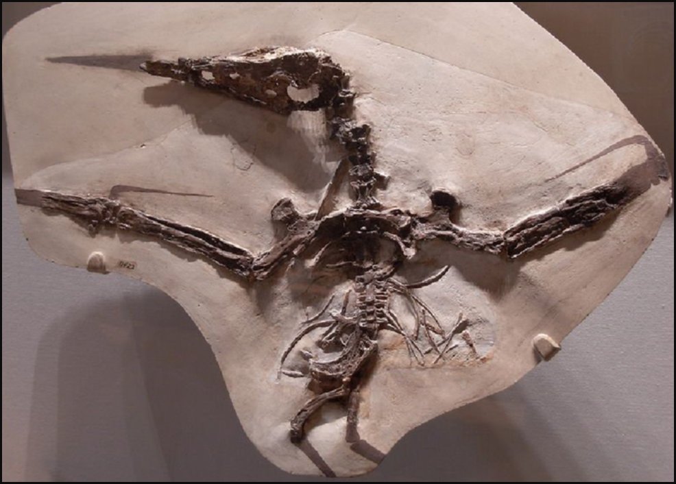 |
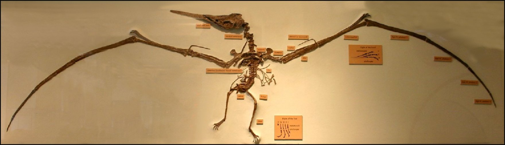
LEFT: A mostly articulated Nyctosaurus specimen (AMNH FR
1716)
collected by H.T. Martin in 1920 from Logan County, Kansas and sold to the
Carnegie Museum in Pittsburg, PA. It was then traded to the AMNH in
1941.
RIGHT: A cast of the same Nyctosaurus specimen reconstructed about 1941 for exhibit in the American Museum of Natural History (AMNH FR 1716). Note that the wings and lower legs are all reconstructed. In this case the wings are wrong because they include the 4 phalange and wing claws. The same specimen is shown in a 1943 article entitled "Flying Reptiles" by Barnum Brown. |
 |
The recent discovery in the Smoky Hill Chalk of western Kansas of two specimens of Nyctosaurus with very large crests is discussed in a paper by Chris Bennett: Bennett, S. C. 2003. New crested specimens of the Late Cretaceous pterosaur Nyctosaurus. Paleontologist Zeitschrift, 77:61-75. (See photographs of the actual specimens here:Chris Bennett web page). Note that these specimens are small (2 m wingspread) and were collected in the lower chalk of Trego County. Based only on their stratigraphic occurrence, it is likely they represent mature males of the earlier smaller species, Nyctosaurus nanus (Marsh, 1881)
 |
LEFT: A cast of the reconstructed skull of one of the Nyctosaurus specimens found and prepared by Kenneth Jenkins from the Smoky Hill Chalk (cast also by
Kenneth Jenkins). Note that this version of the KJ1 specimen was reconstructed to
move the lower jaw upward in articulation, and to remove non-cranial bones. The extremely
large crest on these small flying reptiles raises questions about what it was used for and
how they were able to fly.
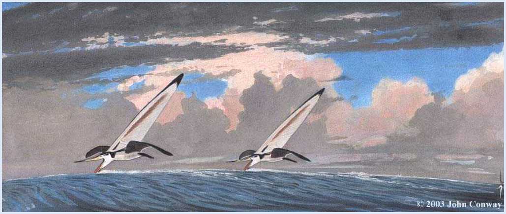
RIGHT: "Skimming Nyctosaurs" painting © 2003 by John Conway. Used with permission of John Conway. (Click on picture to enlarge). While this idea is interesting, only the most mature males would be able to participate in "wind surfing" for prey. |
 |
 |
LEFT: As of 2009, the crested Nyctosaurus (KJ1)
described by Bennett (2003) was on exhibit in the Brazos Valley Museum of
Natural History, in Bryan, Texas. Scale = 10 cm.
RIGHT: The second reported Nyctosaurus (KJ2) with a crest is also on exhibit at the BVMNH. Note that the crest has been reconstructed in this exhibit.. In the original specimen, the crest was broken off near the top of the skull, but was present nearby. While this huge crest must have presented some problems for these individuals, the fact that their remains were found so far from the nearest shore at the time (200 miles or more) means that they were still very capable fliers. |
 |
 |
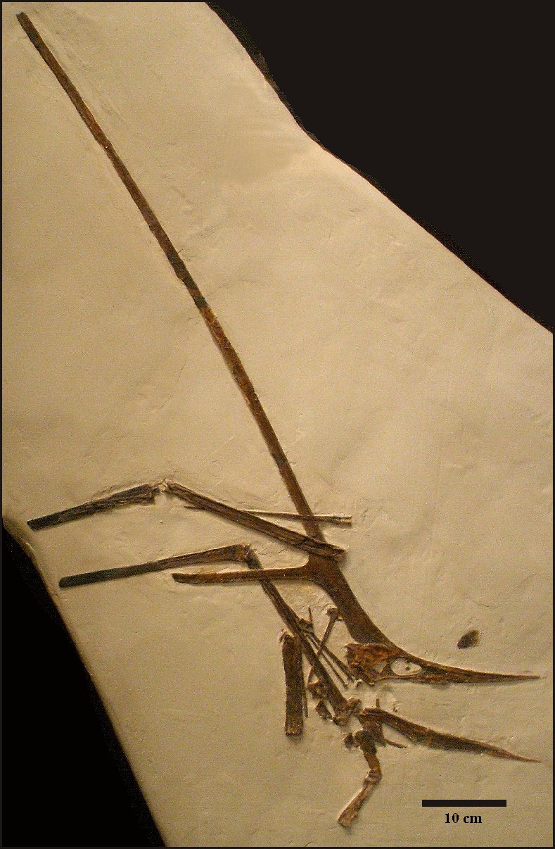
LEFT: The most recently collected partial specimen of Nyctosaurus... probably Nyctosaurus nanus since it was collected in the lower Smoky Hill Chalk of western Gove County, Kansas. Specimen is in a private collection; photos used with permission. |
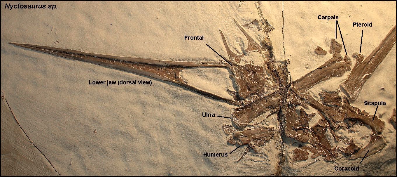 |
LEFT: A closer view of the head of the same specimen, showing an unusual dorsal view of the top of the skull (frontals) and the prefrontals. Note that this specimen shows no evidence of a crest of any sort. (Frontal in dorsal view here). The lower jaw is complete except for a small portion of the distal end. All of the cervical vertebrae are present (four are shown here) |
Pteranodons from the Pierre Shale - The last pteranodons
While most Pteranodon specimens have been collected from the Smoky Hill Chalk Member (Late Coniacian - Early Campanian) in Kansas, there are a significant number specimens (60+) known from the Pierre Shale Group in South Dakota, Wyoming and Kansas (Early to Middle Campanian), including one specimen supposedly collected by Barnum Brown (1904) as stomach contents of a plesiosaur (See Hargrave, 2007, for discussion of Pierre Shale specimens in the Geology Museum at the South Dakota School of Mines). From the specimens that have been found to date, it appears that pteranodons flourished in North America from from about 90 mya to about 80 mya. They may have been around a lot longer but their remains are fragile and not easily preserved in most environmental settings. The Smoky Hill Chalk, deposited far from shore, apparently had the best conditions for the preservation of large numbers of these flying reptiles.
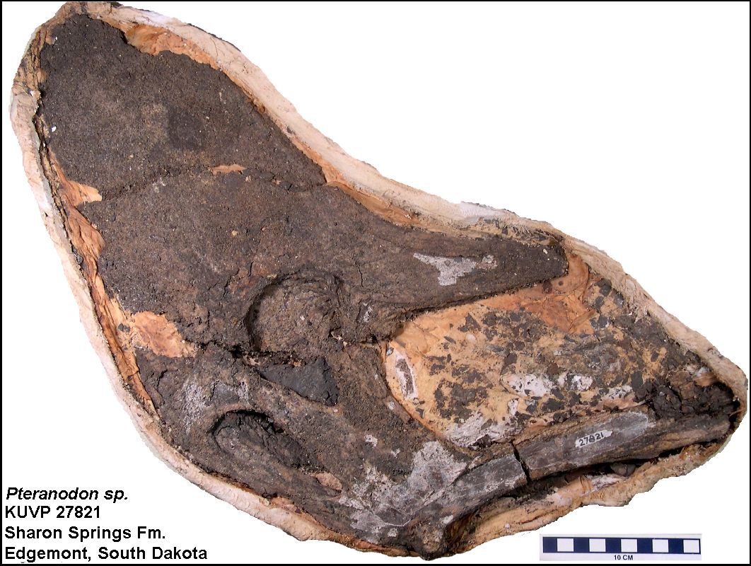 |
LEFT: KUVP 27821 - A partial Pteranodon sp. skull in right lateral view from the Sharon Springs Fm. (formerly the Sharon Springs Member of the Pierre Shale), Edgemont area, southwestern South Dakota (figured by Bennett, 1992, Fig. 4E; Carpenter. 2006, Fig. 14 B). A photo of a Pteranodon bone (KUVP 33564) from the Pierre Shale of western Kansas is shown here Note that Kellner (2010) described this specimen as the holotype of a new species - "Geosternbergia maiseyi” sp. nov. |
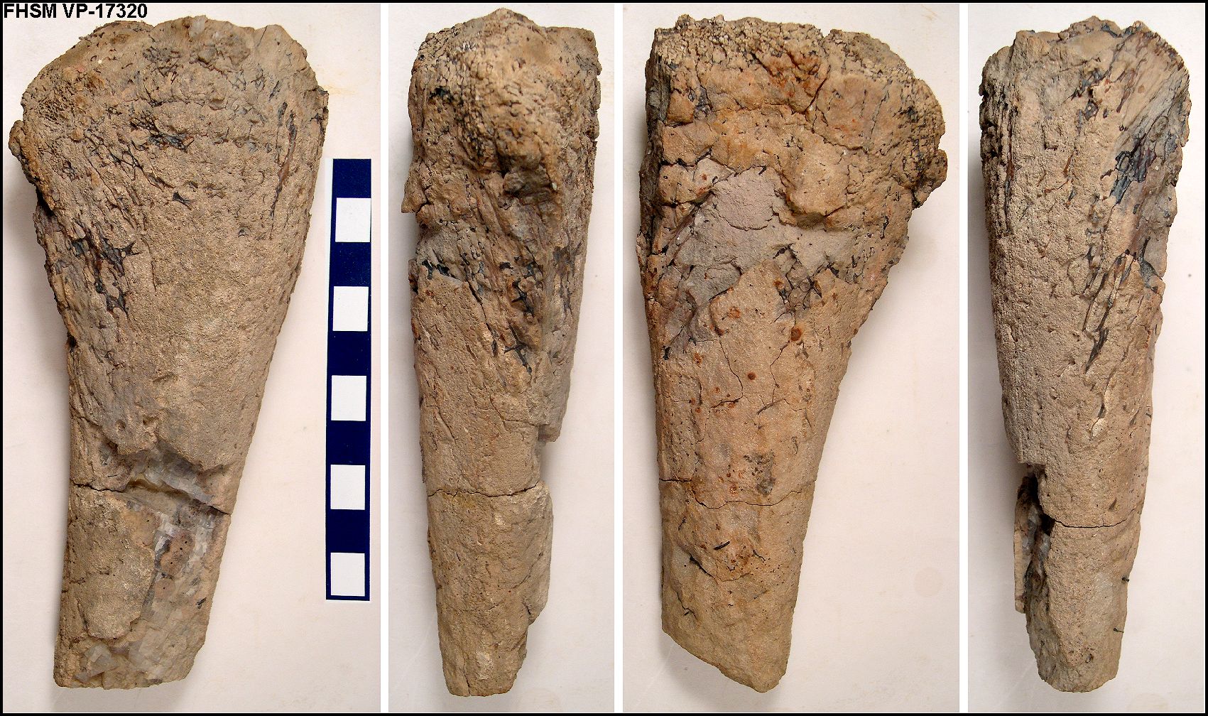 |
LEFT: Four views of an internal cast of the proximal end
of a large Pteranodon longiceps ulna (FHSM VP-17320) discovered by
Pete Bussen in the Sharon Springs Shale, Wallace County, Kansas. Most of
the outer layer of bone has been eroded away, but the internal bone struts
are plainly visible. The bone was apparently filled with a limey sediment
before it could be crushed by the weight of overlying sediments. The
approximate shape of the actual bone is preserved.
RIGHT: the proximal end of the same bone. |
 |
The following chart provides some information regarding where the specimens were collected and in what museum they are curated:
|
Pierre Shale Pteranodon specimens- (Updated from Carpenter dissertation (1996) Number Locality Material Comments AMNH 5803 Loc.
4
plesiosaur Pteranodon bones as stomach contents in concretion in a plesiosaur (AMNH FR 5803) See Brown, 1904 |
Earliest (oldest) pterosaur specimens from Kansas
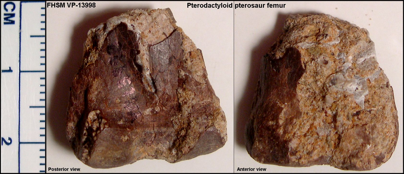 |
LEFT: The distal (lower) end of the right femur of a pterodactyloid pterosaur (FHSM VP-13998) from the basal Lincoln Limestone Member of the Greenhorn Limestone (early Upper Cenomanian in age) - Russell County, Kansas. This specimen is the earliest known pterosaur remains from the state, and came from a flying reptile with a wingspread of about 3.5 m (10 feet) according to Chris Bennett (see Liggett, et al., 2005): "Although the specimen is indistinguishable from Pteranodon from the Niobrara Chalk (Coniacian-early Campanian), it does not exhibit any characters that would enable assignment to a known pterodactyloid taxon." |
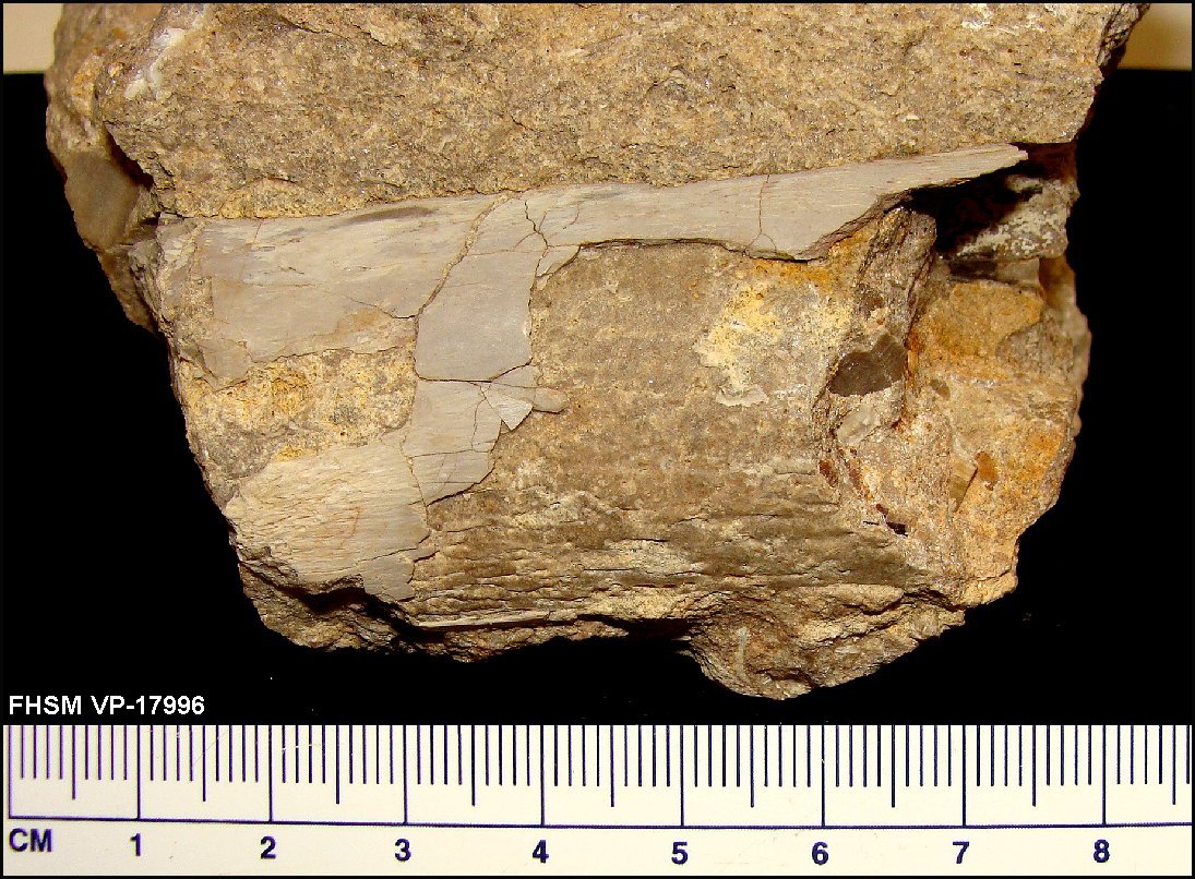 |
LEFT: A second pterosaur bone fragment (FHSM VP-17996) was collected by
Bruce Schumacher in May, 2010, also from the basal Lincoln Limestone Member of the Greenhorn
Limestone (early Upper Cenomanian in age) - Russell County, Kansas. This
appears to be a wing bone, possibly the ulna or radius, from a fairly
large pterosaur. Note that the bone does not appear to be deformed and
apparently had been broken open and filled with sediment before
fossilization. Fragments of inoceramid shell are visible inside the hollow
bone.
RIGHT: An oblique view of the end of the bone fragment, showing how thin the outer layer of bone layer compared to the overall size of the bone. The diameter of this fragment is just over 2.5 cm (~1 in.). The small pits on the surface of the infilling matrix mark the location of the internal struts of the original bone. |
 |
Chris Bennett's Pterosaur webpage HERE
See The Pterosaur Database HERE
Pterosaur Site (Jurassic pterosaurs) HERE
Further reading:
Anonymous. 1872. On two new Ornithosaurians from Kansas. American Journal of Science, Series 3, 3(17):374-375. (Probably written by O. C. Marsh)
Arbour,
V. M. and Currie, P.J.(2011. An istiodactylid pterosaur from the Upper
Cretaceous Nanaimo Group, Hornby Island,
Arbour, V.M. and Currie, P.J. 2011. Corrigendum: an istiodactylid pterosaur from the Upper Cretaceous Nanaimo Group, Hornby Island, British Columbia, Canada. Canadian Journal of Earth Sciences 48:778.
Bennett, S. C. 1987. New evidence on the tail of the pterosaur Pteranodon(Archosauria: Pterosauria). pp. 18-23 In Currie, P. J. and E. H. Koster (eds.), Fourth Symposium on Mesozoic Terrestrial Ecosystems, Short Papers. Occasional Papers of the Tyrrell Museum of Paleontology, #3.
Bennett, S.C. 1990. Inferring stratigraphic position of fossil vertebrates from the Niobrara Chalk of western Kansas. pp. 43-72, In Bennett, S. C. (ed.), Niobrara Chalk Excursion Guidebook, The University of Kansas Museum of Natural History and the Kansas Geological Survey.
Bennett, S.C. 1992. Sexual dimorphism of Pteranodon and other pterosaurs, with comments on cranial crests. Journal of Vertebrate Paleontology 12 p. 422-434.
Bennett, S.C.1993. The ontogeny of Pteranodon and other pterosaurs. Paleobiology 19(1):92-106.
Bennett, S.C. 1994. Taxonomy and systematics of the Late Cretaceous pterosaur Pteranodon (Pterosauria, Pterodactyloidea). Occasional Papers of the Natural History Museum, University of Kansas. 169:1-70.
Bennett ,
Bennett ,
Bennett, S.C. 2000. Inferring Stratigraphic Position of Fossil Vertebrates from the Niobrara Chalk of western Kansas. KansasGeological Survey, Current Research in Earth Sciences, Bulletin 244, part 1. Photos are here
Bennett, S.C. 2000. Pterosaur flight: the role of actinofibrils in wing function. Historical Biology, 14:255-284.
Bennett, S.C. 2000. New information on the skeletons of Nyctosaurus. Journal of Vertebrate Paleontology 20(Supplement to Number 3): 29A. (Abstract)
Bennett, S.C. 2001. The osteology and functional morphology of the Late Cretaceous pterosaur Pteranodon. Part I. General description of osteology. Palaeontographica, Abteilung A, 260:1-112.
Bennett, S.C. 2001.The osteology and functional morphology of the Late Cretaceous pterosaur Pteranodon. Part II. Functional morphology. Palaeontographica, Abteilung A, 260:113-153.
Bennett, S.C. 2003. New crested specimens of the Late Cretaceous pterosaur Nyctosaurus. Paläontologische Zeitschrift, 77:61-75.
Bennett, S.C. 2003. A survey of pathologies of large pterodactyloid pterosaurs. Palaeontology 46(1):185-198.
Bennett, S.C. 2007. Articulation and function of the pteroid bone of pterosaurs. Journal of Vertebrate Paleontology. 27:881-891.
Betts, C. W. 1871. The Yale College expedition of 1870. Harper’s New Monthly Magazine, 43(257):663-671. (Issue of October, 1871)
Bonner, O. W. 1964. An osteological study of Nyctosaurusand Trinacromerum with a description of a new species of Nyctosaurus.Unpub. Masters Thesis, Fort Hays State University, 63 pages.
Brower, J. C. 1983. The aerodynamics of Pteranodonand Nyctosaurus, two large pterosaurs from the Upper Cretaceous of Kansas. Journal of Vertebrate Paleontology 3(2):84-124.
Brower, J.C. 1983. The aerodynamics of Pteranodonand Nyctosaurus, two large pterosaurs from the Upper Cretaceous of Kansas. Journal of Vertebrate Paleontology 3(2):84-124.
Brown, B.1904. Stomach stones and the food of plesiosaurs. Science, 20(501):184-185.
Brown, B. 1943. Flying reptiles. Natural History. 52:104-111.
Brown, G.W. 1978. Preliminary report on an articulated specimen of Pteranodon (Nyctosaurus) gracilis (abstr.). Nebraska Academy of Sciences, Proceedings 88:39.
Brown, G.W. 1986. Reassessment of Nyctosaurus; new wings for an old pterosaur (abstr.). Nebraska Academy of Sciences, Proceedings 96:47.
Brown, G. 1986. Evidence of phalangeal reduction in the wing of the pterosaur Nyctosaurus. Society of Vertebrate Paleontology, Abstracts of the Annual Meeting.
Carpenter, K. 1996. Sharon Springs Member, Pierre Shale (Lower Campanian) depositional environment and origin of its vertebrate fauna, with a review of North American plesiosaurs. Unpub. Ph.D. dissertation, University of Colorado, 251 pp.
Carpenter, K. 2006. Comparative vertebrate taphonomy of the Pembina and Sharon Springs members (Middle Campanian) of the Pierre Shale, Western Interior. Paludicola 5:125-149.
Claessens, L. P. A. M., O’Connor, P.M., and Unwin, D. M. 2009. Respiratory evolution facilitated the origin of pterosaur flight and aerial gigantism. PLoS ONE 4(2): 1-8. (AVAILABLE ONLINE)
Cope,
E.D. 1866. Communication in regard to the Mesozoic sandstone of
Cope,
E.D. 1870. Rhabopelix longispinis,
Cope., pp. 169-175 in Synopsis of
the extinct Batrachia, Reptilia and Aves of
Cope, E. D. 1872. On the geology and paleontology of the Cretaceous strata of Kansas. Annual Report of the U. S. Geological Survey of the Territories 5:318-349 (Report for 1871).
Cope, E. D. 1872. On two new ornithosaurians from Kansas. Proceedings of the American Philosophical Society 12(88):420-422.
Cope, E. D. 1874. Review of the vertebrata of the Cretaceous period found west of the Mississippi River. U. S. Geological Survey of the Territories Bulletin 1(2):3-48.
Cope, E. D. 1875. The vertebrata of the Cretaceous formations of the West. Report, U. S. Geological Survey of the Territories (Hayden). 2:302 p, 57 pls.
Currie, P.J., and Padian, K. 1983. A new pterosaur record from the Judith River (Oldman) Formation of Alberta. Journal of Paleontology 57(3):599-600.
Currie, P.J. and Russell D.A. 1982. A giant pterosaur (Reptilia: Archosauria) from the Judith River Oldman Formation of Alberta. Canadian Journal of Earth Science. 19: 894-897.
Eaton, G. F. 1903. The characters of Pteranodon. American Journal of Science, ser. 4, 16(91):82-86, pl. 6-7.
Eaton, G. F. 1904. The characters of Pteranodon(second paper). American Journal of Science, ser. 4, 17(100):318-320, pl. 19-20.
Eaton, G. F. 1908. The skull of Pteranodon. Science (n. s.) XXVII 254-255.
Eaton, G. F. 1910. Osteology of Pteranodon. Memoirs of the Connecticut Academy of Arts and Sciences, 2:1-38, pls. i-xxxi.
Edinger, T. 1927. Das Gehirn der Pterosaurier. Zeitschrift Anat. Entwicklungsgeschichte 83:105-112. ["The brain of the pterosaur" - In German]
Elgin, R.A., Grau, C.A., Palmer, C., Hone, D.W.E., Greenwell, D. and Benton, M.J. 2008. Aerodynamic characters of the cranial crest in Pteranodon. Zitteliana B28:167-174.
Elgin, R.A., Hone, D.W.E., and Frey, E. 2011. The extent of the pterosaur flight membrane. Acta Palaeontologica. Polonica 56(1):99-111.
Everhart, M. J. and Everhart, P. 1999. An early occurrence of Pteranodon sternbergi from the Smoky Hill Member (Late Cretaceous) of the Niobrara Chalk in western Kansas. Kansas Academy of Science, Abstracts of the 131st Annual Meeting 18:27.
Frey, E., Buchy, M.-C. Martill and D.M. 2003. Middle- and bottom-decker Cretaceous pterorsaurs: unique designs in active flying vertebrates. in Buffetaut, E. and Mazin, J.-M. (eds.), Evolution and Paleobiology of Pterosaurs. Geological Society, London, Special Publications 217:267-274.
Frey, E. Martill, D.M. and Buchy, M.-C. 2003. A new species of tapejarid pterosaur with soft-tissue head crest. in Buffetaut, E. and Mazin, J.-M. (eds.), Evolution and Paleobiology of Pterosaurs. Geological Society, London, Special Publications 217:65-72.
Frey, E., Buchy, M.-C., Stinnesbeck,
W., González- González, A. and Stefano, A. 2006. Muzquizopteryx
coahuilensis n.g., n.sp., a nyctosaurid pterosaur with soft tissue
preservation from the Coniacian (Late Cretaceous) of northeast
Habib, M. 2008. Comparative
evidence for quadrupedal launch in pterosaurs. Pp 159-166 in Buffetaut E., and
D.W.E. Hone (eds.), Wellnhofer Pterosaur Meeting: Zitteliana B28.
Hargrave, J.E. 2007. Pteranodon (Reptilia: Pterosauria): Stratigraphic distribution and taphonomy in the lower Pierre Shale Group (Campanian), western South Dakota and eastern Wyoming. p.215-225 in Martin, J.E. and Parris, D.C. (eds.), The Geology and Paleontology of the Late Cretaceous Marine Deposits of the Dakotas. Geological Society of America, Special Paper 427.
Harksen, J. C. 1966. Pteranodon sternbergi, a new fossil pterodactyl from the Niobrara Cretaceous of Kansas. Proceedings South Dakota Academy of Science 45:74-77.
Henderson, M.D. and Peterson, J.E. 2006. An Azhdarchid pterosaur cervical vertebra from the Hell Creek Formation (Maastrichtian) of southeastern Montana. Journal of Vertebrate Paleontology 26(1):192–195.
Henderson, D.M. 2010. Pterosaur body mass estimates from three-dimensional mathematical slicing. Journal of Vertebrate Paleontology 30(3):768-785.
Huene,
F. von. 1921. Reptilian and stegocephalian remains from the Triassic of
Humphries, S., Bonser, R.H.C., Witton, M.P., and Martill, D.M. 2007. Did pterosaurs feed by skimming? Physical modeling and anatomical evaluation of an unusual feeding method. PLoS Biology 5(8):1648-1655.
Kellner,
A.W.A. 2010. Comments on the Pteranodontidae (Pterosauria, Pterodactyloidea)
with description of two new species. Anais da Academa Brasileira de Cięncias
82(4):1063-1084.
Lane, H. H. 1946. A survey of the fossil vertebrates of Kansas, Part III, The Reptiles, Kansas Academy Science, Transactions 49(3):289-332, figs. 1-7.
Liggett, G. A., S. C. Bennett, K. Shimada and J. Huenergarde. 1997. A Late Cretaceous (Cenomanian) fauna in Russell county, KS. Kansas Academy of Science, Transactions Abstracts. 16:26
Liggett, G. A., K. Shimada, S. C. Bennett and B. Schumacher. 2005. Cenomanian (Late Cretaceous) reptiles from northwestern Russell County, Kansas. PaleoBios 25(2): 9-17.
Lü, J., Unwin, D.M., Deeming, D.C., Jin, X., Liu, Y. and Ji, Q. 2011. An egg-adult association, gender, and reproduction in pterosaurs. Science 331(6015):321-324. (January 21, 2011)
Lucas,
F.A. 1902. The greatest flying creature, the great Pterodactyl Ornithostoma.
Report of the Smithsonian Institution 1901:654-659.
Marsh, O. C. 1871. Scientific expedition to the Rocky Mountains. American Journal of Science ser. 3, 1(6):142-143.
Marsh, O. C. 1871. Notice of some new fossil reptiles from the Cretaceous and Tertiary formations. American Journal of Science, Series 3, 1(6):447-459.
Marsh, O. C. 1871. Note on a new and gigantic species of Pterodactyle. American Journal of Science, Series 3, 1(6):472.
Marsh, O. C. 1872. Discovery of additional remains of Pterosauria, with descriptions of two new species. American Journal of Science, Series 3, 3(16) :241-248.
Marsh, O. C. 1876. Notice of a new sub-order of Pterosauria. American Journal of Science, Series 3, 11(65):507-509.
Marsh, O. C. 1876. Principal characters of American pterodactyls. American Journal of Science, Series 3, 12(72):479-480.
Marsh, O. C. 1881. Note on American pterodactyls. American Journal of Science, Series 3, 21(124):342-343.
Marsh, O. C.. 1882. The wings of Pterodactyles. American Journal of Science, Series 3, 23(136):251-256, pl. III.
Marsh, O. C. 1884. Principal characters of American Cretaceous pterodactyls.
Part I. The skull of Pteranodon. American Journal of Science, Series 3,
27(161):422-426, pl. 15.
Mateer, N.J. 1975. A study of Pteranodon. Bulletin of the Geological Institutions of Uppsala 6:23-33.
Matthew, W.D. 1916. A reptilian aëronaut; a new
skeleton of the giant flying reptile of the Cretaceous Period.
Matthew, W.D. 1920. Flying Reptiles. Natural History 20:73-81.
Miller, H. W. 1971. The taxonomy of the Pteranodonspecies from Kansas. Kansas Academy of Science, Transactions 74(1):1-19.
Miller, H. W. 1971. A skull of Pteranodon (Longicepia)longiceps Marsh associated with wing and body parts. Kansas Academy of Science, Transactions 74(10):20-33.
Miller, H.W. 1978. Geosternbergia, a new name for SternbergiaMiller, 1972: Non Paulo Couto 1970; non Jordon, 1925. Journal of Paleontology 52(1):194.
Myers,
T.S. 2010. Earliest occurrence of the Pteranodontidae (Archosauria: Pterosauria)
in North America: New material from the Austin Group of Texas. Journal of Paleontology 84(6):1071-1081.
Myers, T.S. 2010. A new ornithocherid pterosaur from the Upper Cretaceous (Cenomanian-Turonian) Eagle Ford Group of Texas. Journal of Vertebrate Paleontology 30(1):280–287.
Padian, K. 1983. A functional analysis of flying and walking in pterosaurs. Paleobiology 9(3):218-239.
Padian, K. 1984. A large pterodactyloid pterosaur from the Two Medicine Formation (Campanian) of Montana. Journal of Vertebrate Paleontology 4(4):516-524.
Padian, K., and Smith, M. 1992. New light on Late Cretaceous pterosaur material from Montana. Journal of Vertebrate Paleontology 12(1): 87-92.
Russell, D.A. 1988. A check list of North American marine cretaceous vertebrates including fresh water fishes, Occasional Paper of the Tyrrell Museum of Palaeontology, (4):57.
Schoch, R.M. 1984. Notes on the type specimens of Pteranodon and Nyctosaurus (Pterosauria, Pteranodontidae) in the Yale Peabody Museum Collections. Postilla 194:1-23.
Schultze, H.-P., L. Hunt, J. Chorn and A. M. Neuner. 1985. Type and figured specimens of fossil vertebrates in the collection of the University of Kansas Museum of Natural History, Part II. Fossil Amphibians and Reptiles. Miscellaneous Publications of the University of Kansas Museum of Natural History 77:66 pp.
Schwimmer, D. R. Padian, K., and Woodhead, A.B. 1985. First pterosaur records from Georgia: Open marine facies, Eutaw Formation (Santonian). Journal of Paleontology 59(3):674-676.
Seeley, H. G. 1870. The Ornithosauria: an elementary study of the bones of pterodactyles. Deighton, Bell, and Co., Cambridge, xviii + 135 pp.
Seeley, Harry G. 1871. Additional evidence of the structure of the head in ornithosaurs from the Cambridge Upper Greensand; being a supplement to "The Ornithosauria." The Annals and Magazine of Natural History, Series 4, 7:20-36, pls. 2-3. (Discovery of toothless pterosaurs in England)
Shor, E. N. 1971. Fossils and flies; The life of a compleat scientist - Samuel Wendell Williston, 1851-1918, University of Oklahoma Press, 285 pp.
Sneyd, A.D., Bundock, M.S. and Reid. D. 1982. Possible effects of wing flexibility on the aerodynamics of Pteranodon. The American Naturalist 120(4):455-477.
Stein, R.S. 1975. Dynamic analysis of Pteranodon ingens: A reptilian adaptation to flight. Journal of Paleontology 49(3):534-548.
Sternberg, C. H. 1990. The life of a fossil hunter, Indiana University Press, 286 pp. (Originally published in 1909 by Henry Holt and Company)
Sternberg, G.F. and M.V. Walker. 1958. Observation of articulated limb bones of a recently discovered Pteranodon in the Niobrara Cretaceous of Kansas. Kansas Academy of Science, Transactions, 61(1):81-85.
Stewart, J. D. 1990. Niobrara Formation vertebrate stratigraphy. pp. 19-30 in Bennett, S. C. (ed.), Niobrara Chalk Excursion Guidebook, The University of Kansas Museum of Natural History and the Kansas Geological Survey.
Stinnesbeck, W., Ifrim, C., Schmidt, H.,
Rindfleisch, A., Buchy, M.-C., Frey, E., González-González, A.H., Vega, F.J.
Cavin, L., Keller, G. and Smith, K.T. 2005. A new lithographic limestone deposit
in the Upper Cretaceous Austin Group at El Rosario,
Vullo, R., Buffetaut, E. and Everhart, M.J. 2012. Reappraisal of Gwawinapterus beardi from the Late Cretaceous of Canada: A saurodontid fish, not a pterosaur. Journal of Vertebrate Paleontology 32(5):1198-1201.
Wang, X. and Z. Zhou. 2004. Pterosaur embryo from the Early Cretaceous. Nature 429:621.
Webb, W. E. 1872. Buffalo Land - An authentic account of the discoveries, adventures, and mishaps of a scientific and sporting party in the Wild West. Hubbard Bros., Philadelphia, 503 pp.
Wellnhoffer, P. 1978. Pterosauria. Handbuch der Paläeoherpetologie, Tiel 19, x+82 pp. Gustav Fisher Verlag, Stuttgart.
Wellnhofer, P. 1991. The illustrated encyclopedia of pterosaurs. Crescent Books, New York, 192 pp.
Williston,
S.W. 1885. Über Ornithocheirus hilsensisKoken. Zoologische Anzeiger 8:628-629.
Williston, S.W. 1891. The skull and hind extremity of Pteranodon. American Naturalist 25(300):1124-1126.
Williston, S.W. 1892. Kansas pterodactyls. Part I. Kansas University Quarterly 1:1-13, pl. i.
Williston, S.W. 1893. Kansas pterodactyls. Part II. Kansas University Quarterly 2:79-81, with 1 fig.
Williston, S.W. 1895. Note on the mandible of Ornithostoma. Kansas University Quarterly 4:61.
Williston, S.W. 1896. On the skull of Ornithostoma. Kansas University Quarterly 4(4):195-197, with pl. i.
Williston, S.W. 1897. Restoration of Ornithostoma (Pteranodon). Kansas University Quarterly 6:35-51, with pl. ii.
Williston, S.W. 1902. On the skeleton of Nyctodactylus, with restoration. American Journal of Anatomy. 1:297-305.
Williston, S.W. 1902. On the skull of Nyctodactylus, an Upper Cretaceous pterodactyl. Journal of Geology, 10:520-531, 2 pls.
Williston, S.W. 1902. Winged reptiles. Pop. Science Monthly 60:314-322, 2 figs.
Williston, S.W. 1902. Review of Seeley's Dragons of the Air. Science 15(367):67-68.
Williston, S.W. 1903. On the osteology of Nyctosaurus (Nyctodactylus), with notes on American pterosaurs. Field Mus. Publ. (Geological Ser.) 2(3):125-163, 2 figs., pls. XL-XLIV.
Williston, S.W. 1904. The fingers of pterodactyls. Geology Magazine, Series 5, 1(2): 5:59-60.
Williston, S.W. 1910. A mounted skeleton of Platecarpus. Journal of Geology 18(6):537-541, 1 fig. (Notes that he has seen evidence of a fibula in Pteranodon)
Williston, S.W. 1911 The wing-finger of pterodactyls, with restoration of Nyctosaurus. Journal of Geology. 19:696–705.
Williston, S.W. 1912. A review of G. F. Eaton's "Osteology of Pteranodon." Journal of Geology. 20:288.
Wiman, C. 1920. Some reptiles from the Niobrara Group in Kansas. Bulletin of the Geological Institution of the University of Upsala, Upsala, 18:9-18.
Witton,
M.P. 2010. Pteranodon and beyond: the
history of giant pterosaurs from 1870 onwards. pp. 313-323 in Moody, R.T.J.,
Buffetaut, E., Naish, D. and Martill, D.M. (eds.), Dinosaurs and Other Extinct Saurians: A Historical Perspective.
Geological Society,
Witton,
M.P. and Habib, M.B. 2010. On the size and flight diversity of giant pterosaurs,
the use of birds as pterosaur analogues and comments on pterosaur flightlessness.
PLoS One 5(11):1-18.
Witton, M.P. and Naish, D. 2008. A reappraisal of Azhdarchid pterosaur functional morphology and paleoecology. PLoS Biology 3(5):1-16.

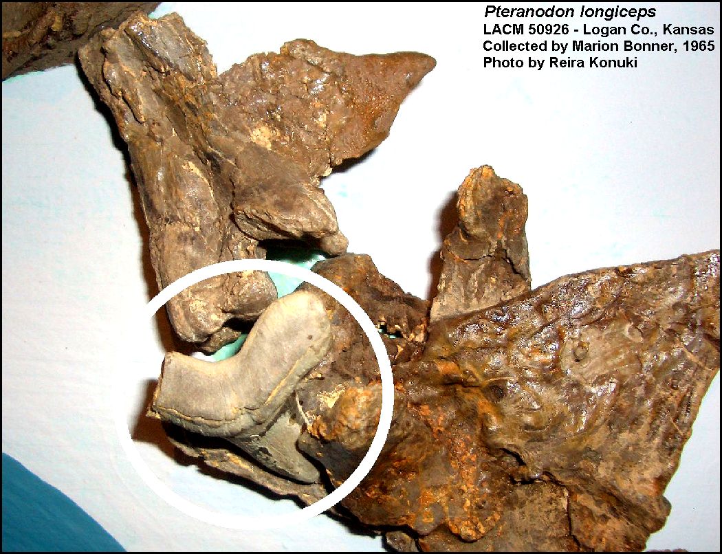
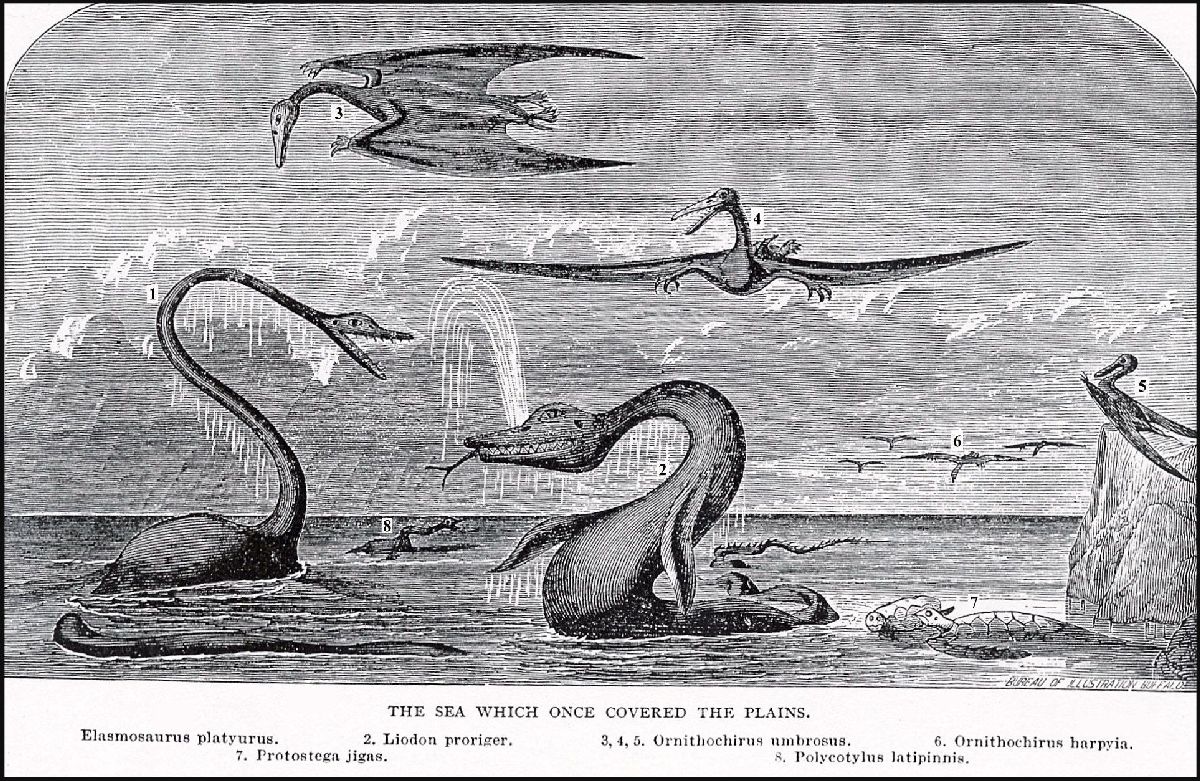
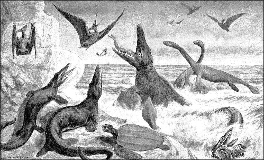
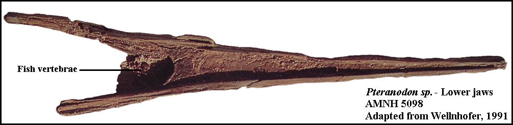
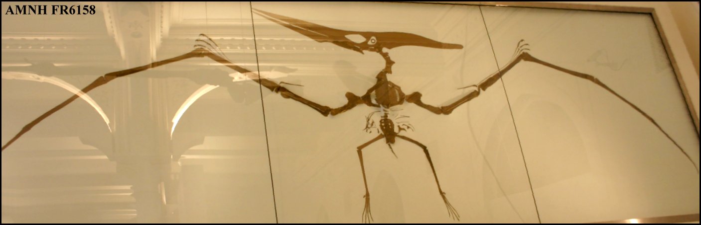
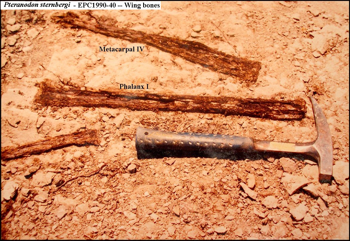
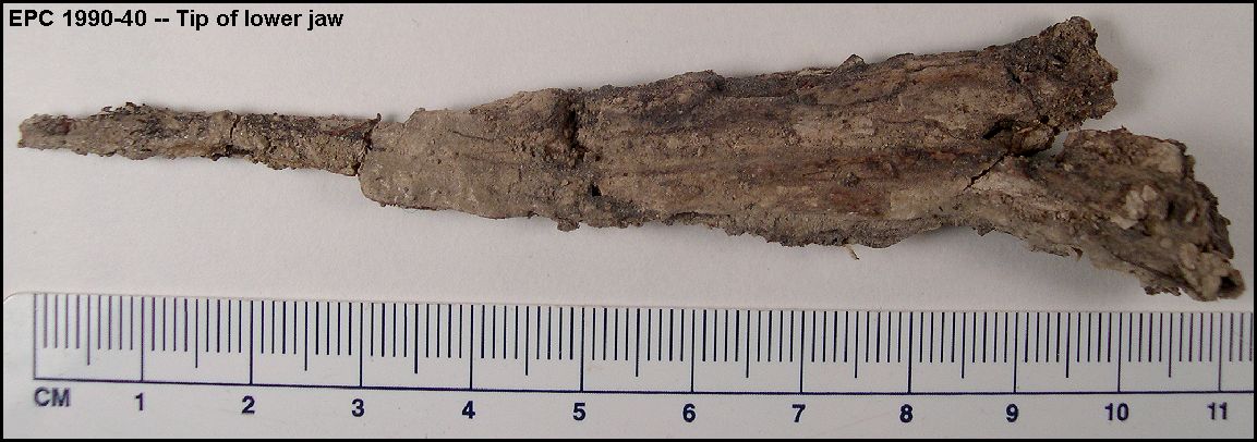
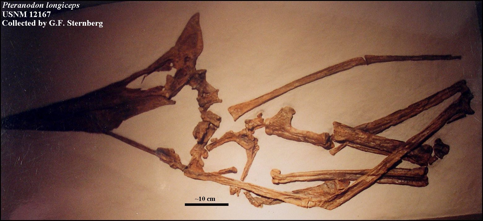
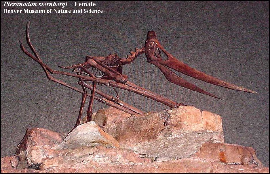
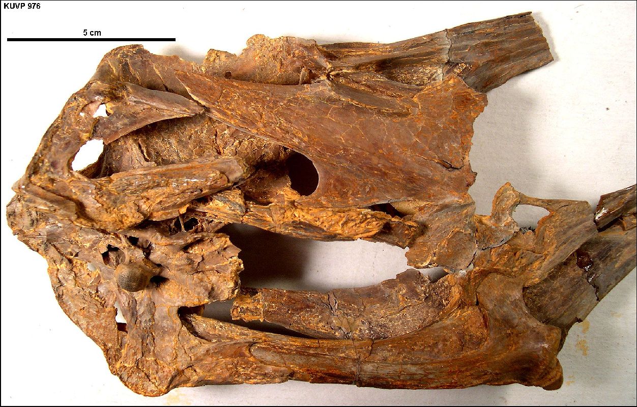
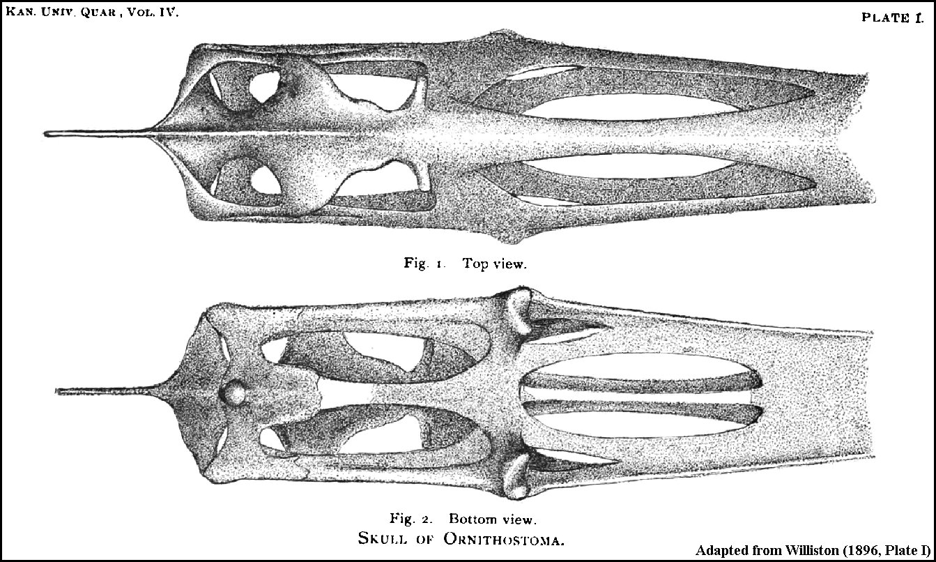
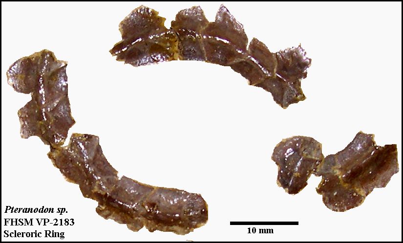
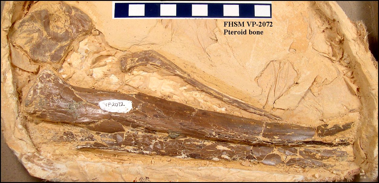
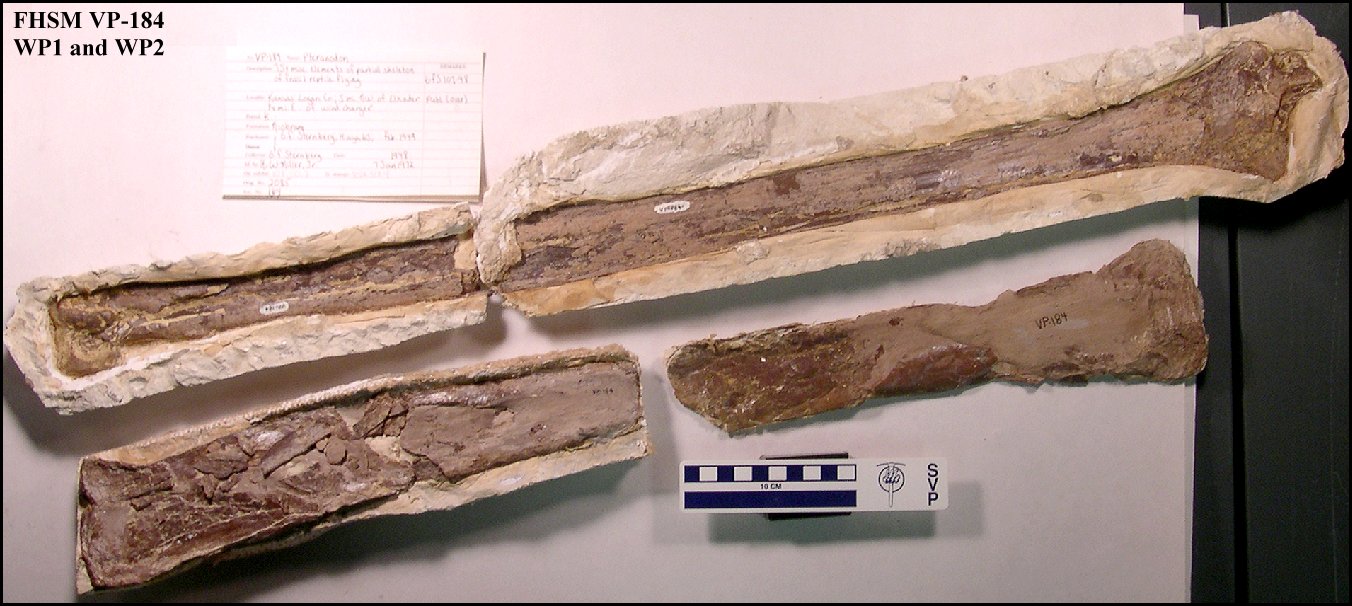
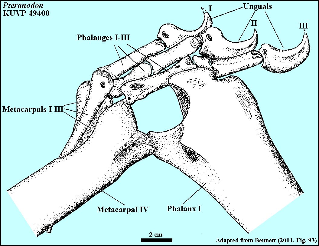
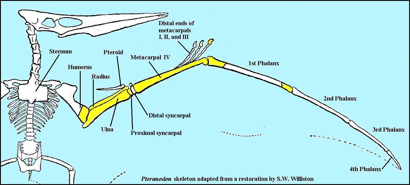
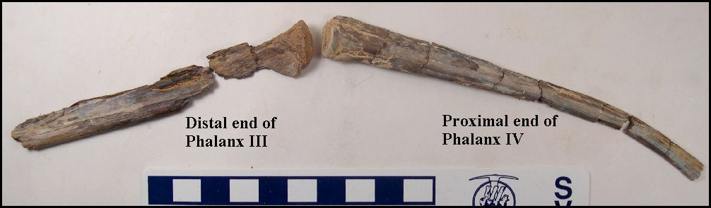
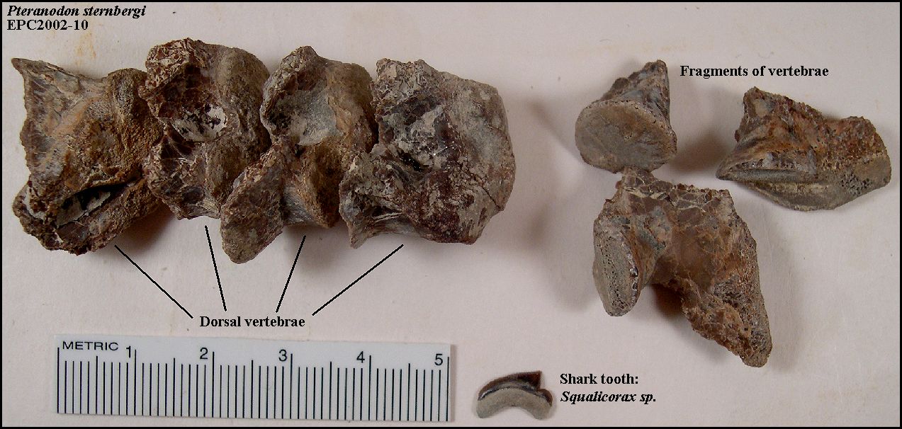
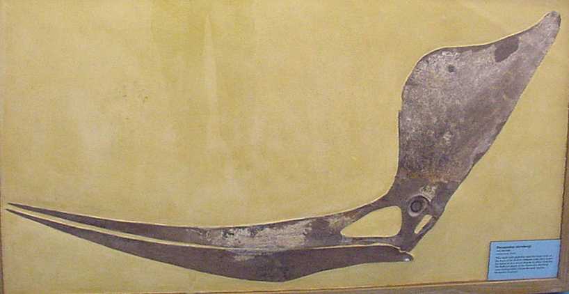
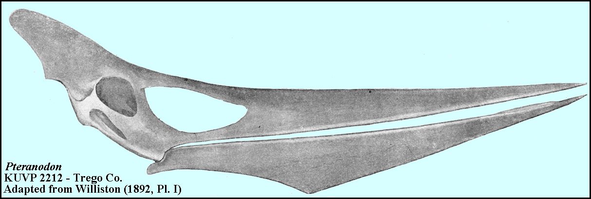
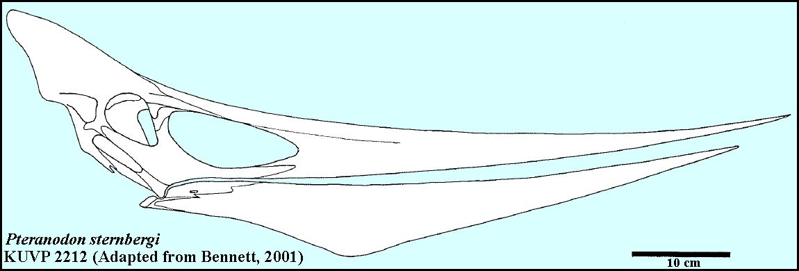
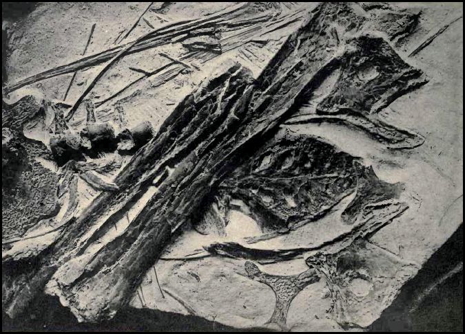
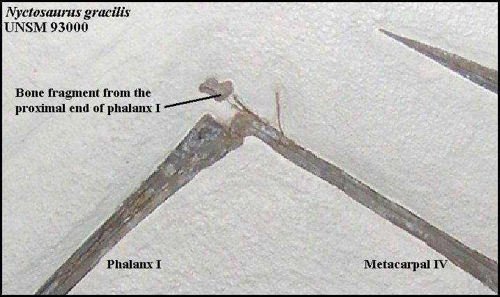
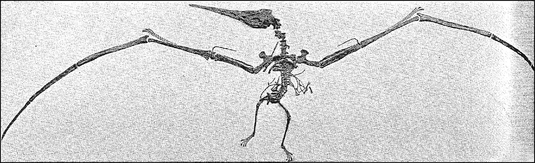
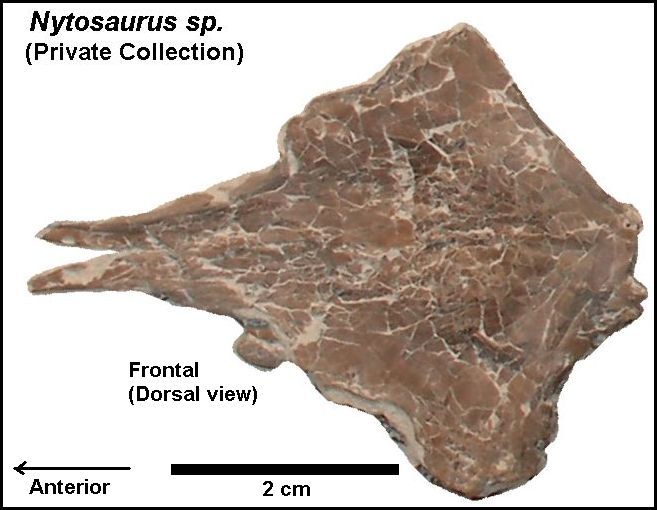
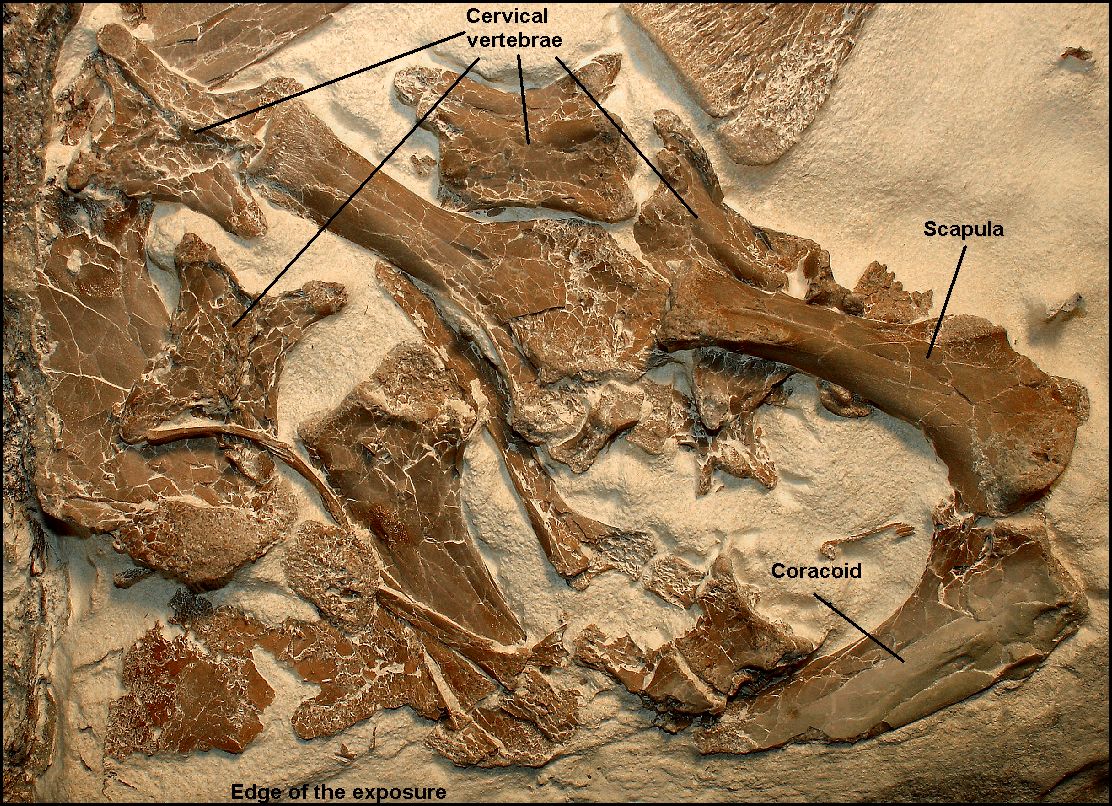