Click on the Thumbnail |
Specimen Description |
Specimen Number / I.D. |
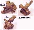  |
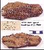
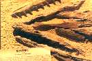
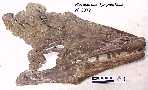
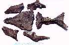
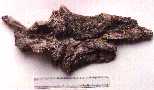
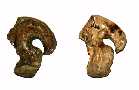
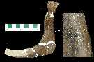
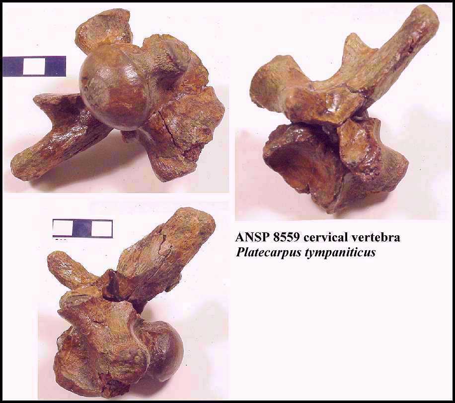
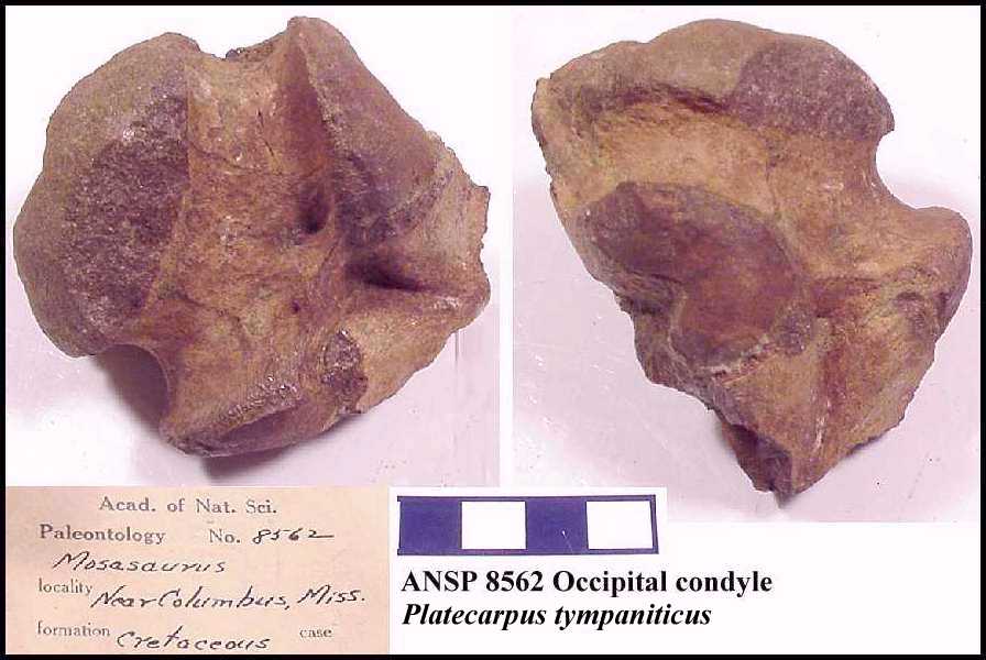
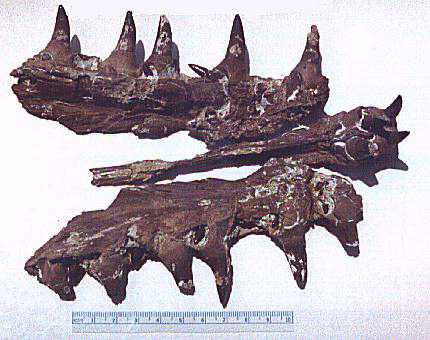
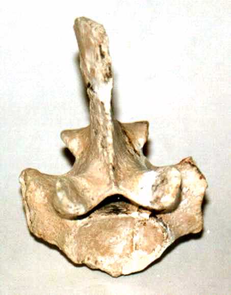
Left: Three views of a cervical
vertebra from the type specimen of Platecarpus tympaniticus Cope
1869, found near
Columbus, Mississippi. Right: A fragment of the basio-occipital of the skull of the type
specimen of Platecarpus tympaniticus Cope. Note per Konishi and Caldwell (2007),
this was the only known specimen of P. tympaniticus. Now (2010)
it's the type species. |
ANSP 8559 and ANSP 8562; Type specimen,
Academy of Natural Sciences of Philadelphia. |
  |
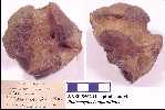
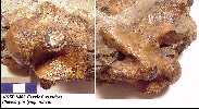
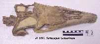
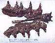
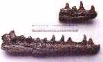
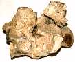
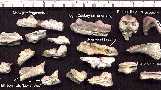
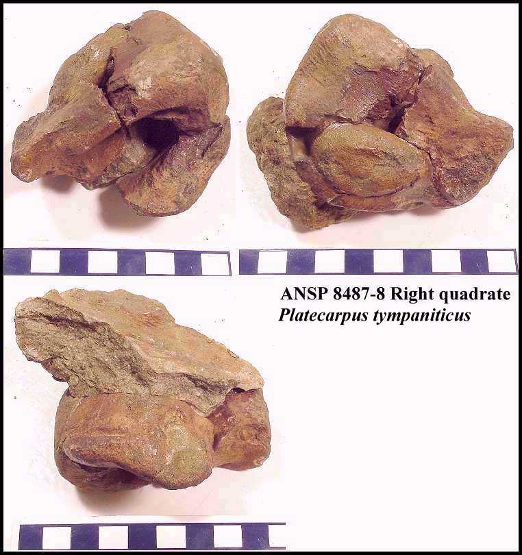
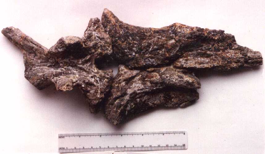
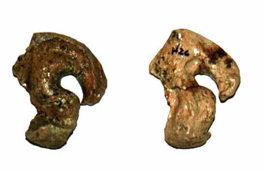
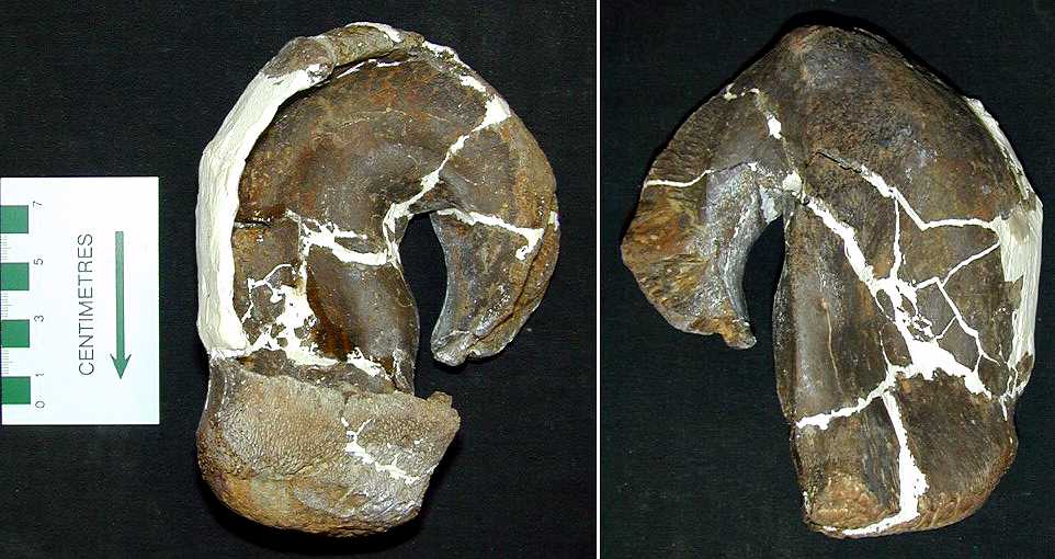
Left: A fragment of a vertebra from the
type specimen. Right: Three views of the right quadrate from the type specimen. Note that
even though the specimen numbers change, these six bones are all part of the same
mosasaur. |
ANSP 8558 and ANSP 8487; Type specimen,
Academy of Natural Sciences of Philadelphia. |
  |
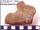
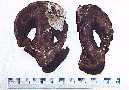
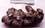
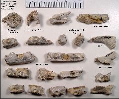
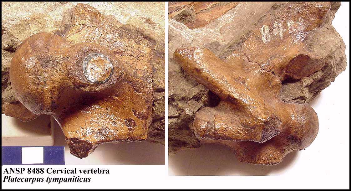
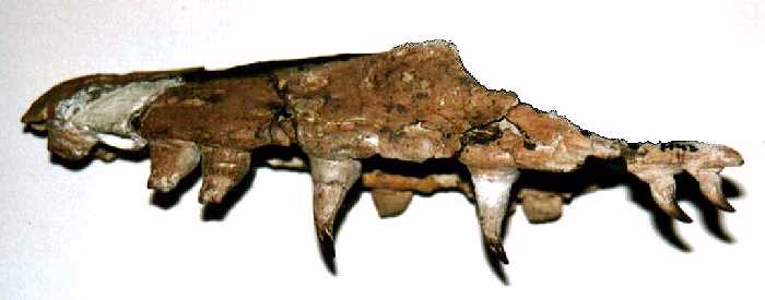
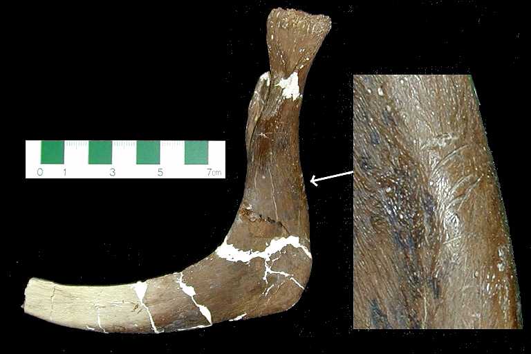
Left: A portion of the lower jaw of the
type specimen. Right: Another cervical vertebra from the type specimen, still in matrix. |
ANSP 8484 and ANSP 8488; Type specimen,
Academy of Natural Sciences of Philadelphia. |
 |
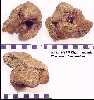
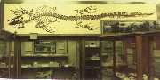
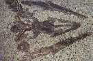
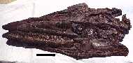
A picture of a Kansas Platecarpus tympaniticus
specimen in
the Tübinger Museum in Germany. Collected by Charles Sternberg
from the Smoky Hill Chalk. |
Natural History Museum, University of Tübingen. From von Huene, 1919. |
 |
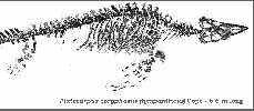
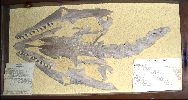
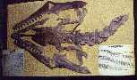
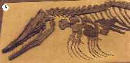
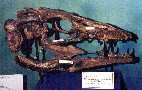
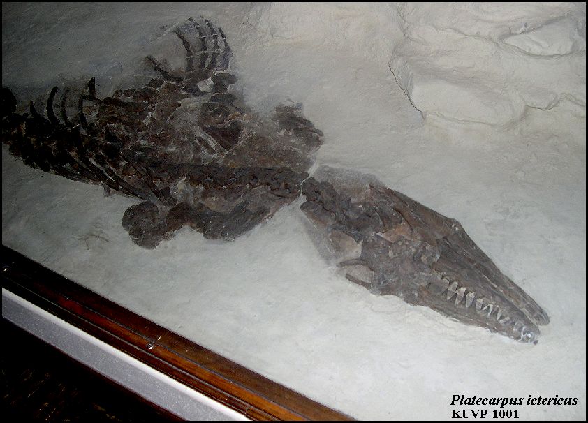
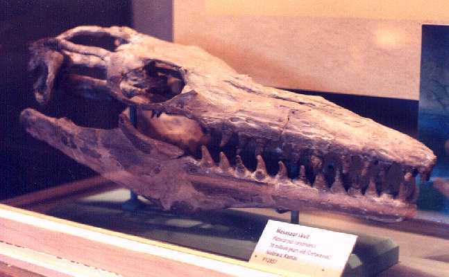
Platecarpus tympaniticus
(KUVP 1001) -
Nearly complete specimen (missing the tail), that preserves calcified cartilage, including
the trachea, skin and scale impressions and stomach contents. |
The University of Kansas, Museum of Natural History |
 |
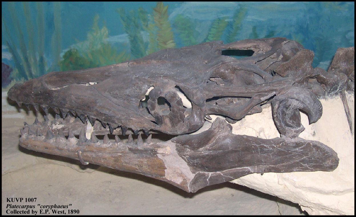
Platecarpus 'coryphaeus' (KUVP
1007), left lateral view of the skull. This specimen was collected by E.P. West in 1890. P.
coryphaeus is also a junior synonym of P. tympaniticus
Cope 1869. |
The University of Kansas, Museum of Natural History |
 |
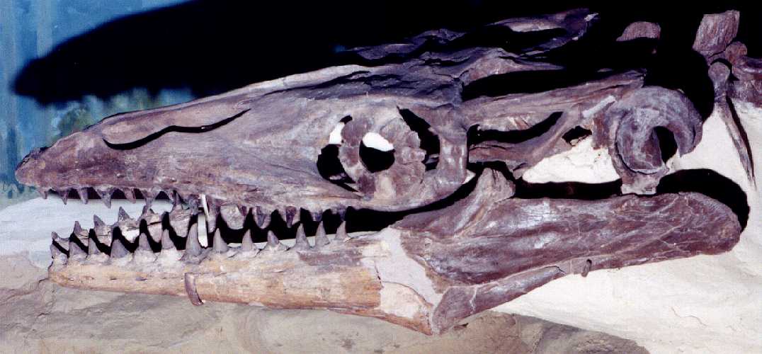
Another left lateral view of the specimen above.
Slightly different angle and lighting. |
The University of Kansas, Museum of Natural History |
 |
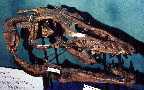
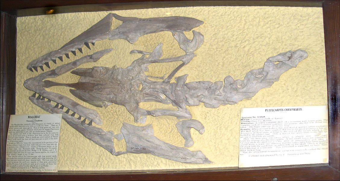
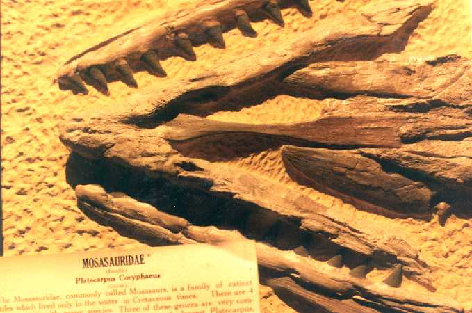
Platecarpus tympaniticus
skull, dorsal view of
skull, lower jaws and cervical vertebrae. Specimen collected and prepared by G. F.
Sternberg. |
Fick Fossil and
History Museum, Oakley, KS |
 |
Platecarpus tympaniticus
skull, dorsal view of
frontal and parietal |
Fick Fossil and History Museum, Oakley, KS |
 |
Platecarpus tympaniticus
skull, dorsal view of premaxilla, and both maxillaries |
Fick Fossil and History Museum, Oakley, KS |
 |
Platecarpus tympaniticus
skull and cervical
vertebrae, slightly different angle and better lighting. |
Fick Fossil and History Museum, Oakley, KS |
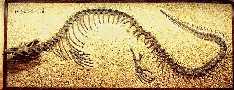 |
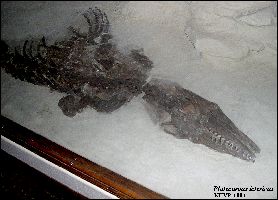
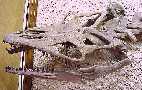
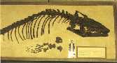
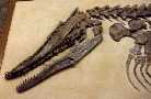
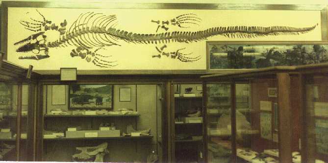
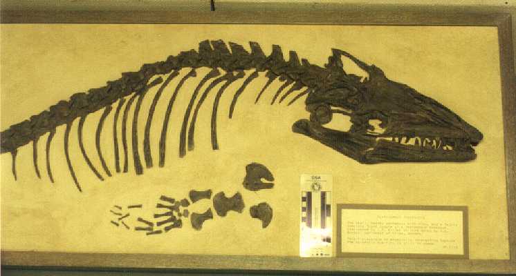
Platecarpus tympaniticus
- A nearly complete
skeleton from Gove County, KS (Smoky Hill Chalk). This specimen was found by a group
of teachers on a field trip. |
Emporia State University Johnston Geology Museum,
Emporia, Kansas |
 |
NEW 2/2000 Platecarpus tympaniticus. Additional
pictures here: back of skull 1, back of skull 2, and back
of skull 3. |
Emporia State University Johntson Geology Museum,
Emporia, Kansas |
 |
NEW 2/2000 Platecarpus tympaniticus. Additional
pictures here: skull 2, skull
and neck 3, and skull 4. |
Emporia State University Johnston Geology Museum,
Emporia, Kansas |
 |
NEW 2/2000 Platecarpus tympaniticus. Additional
pictures here: muzzle, neck
and tail vertebrae. |
Emporia State University Johnston Geology Museum,
Emporia, Kansas |
 |
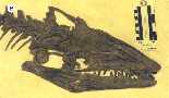
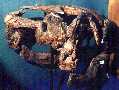
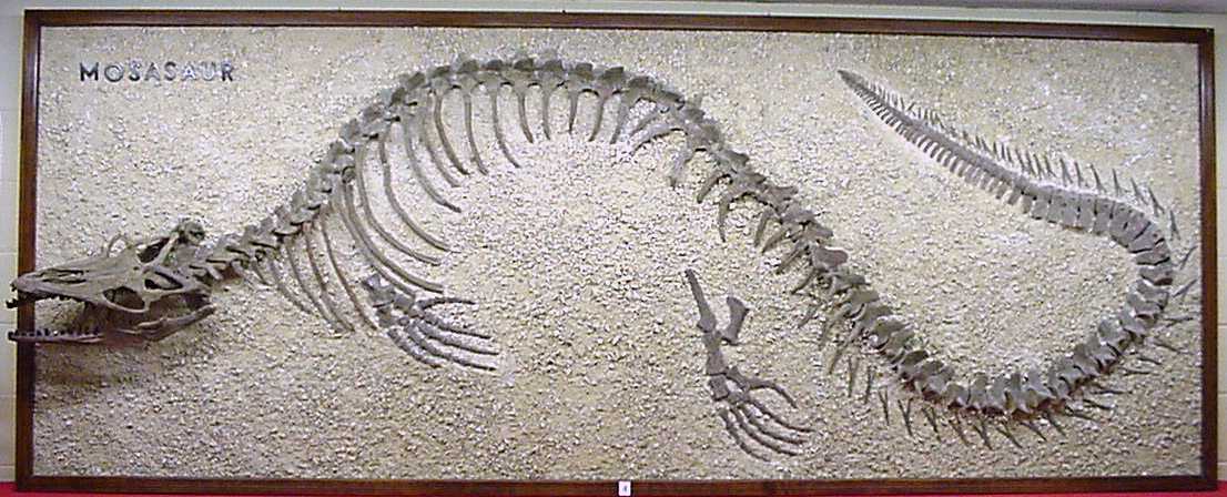
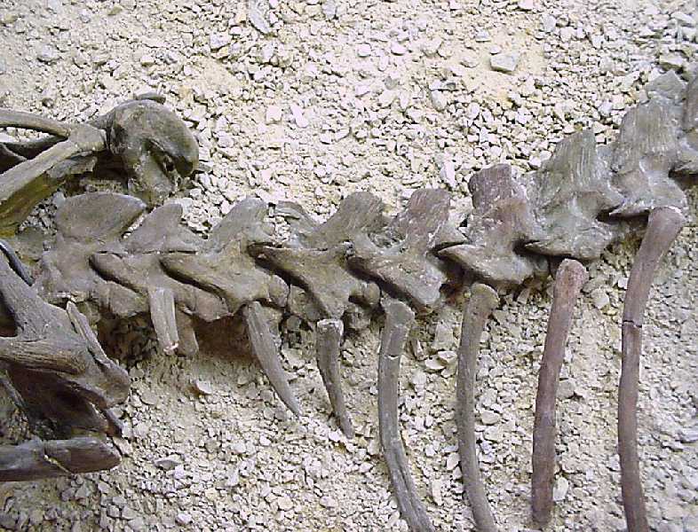
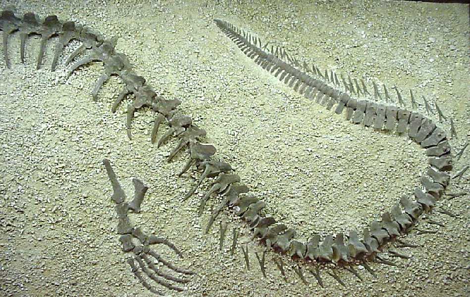
Platecarpus tympaniticus, approximately 6
meters (20 feet) in length, complete skeleton. Although displayed for years in the
old museum, this specimen is now in storage. A recent examination showed a number of previously unnoticed pathologies. |
Sternberg
Museum VP-322 |
 |
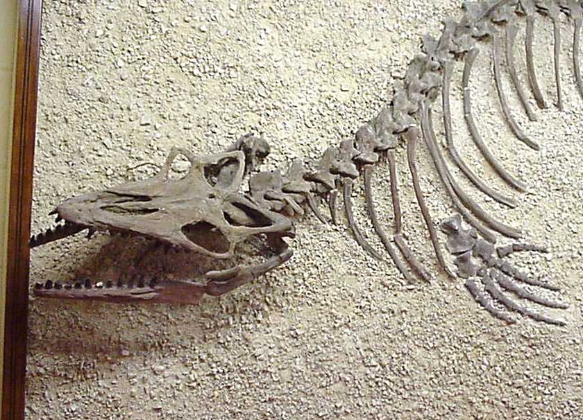
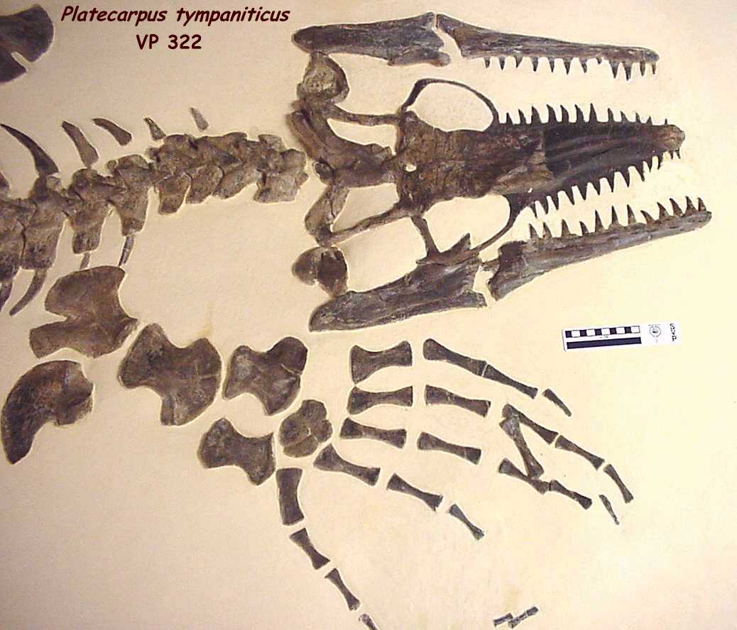
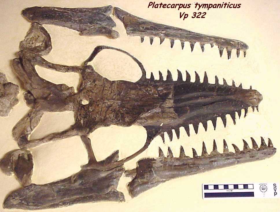
Platecarpus tympaniticus
skull from the
above specimen, dorsal view. NEW 2/2000 - For close-ups of the skull, click on the
following: skull1 and skull 2. |
Sternberg Museum VP-322 |
 |
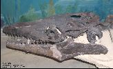
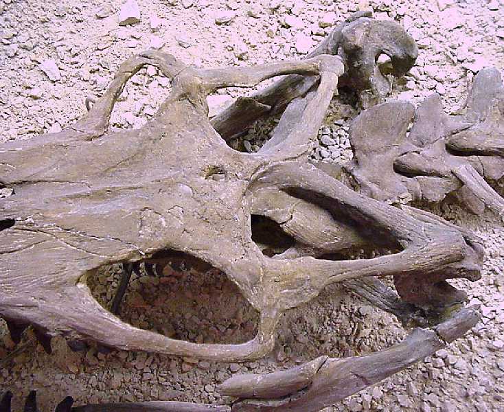
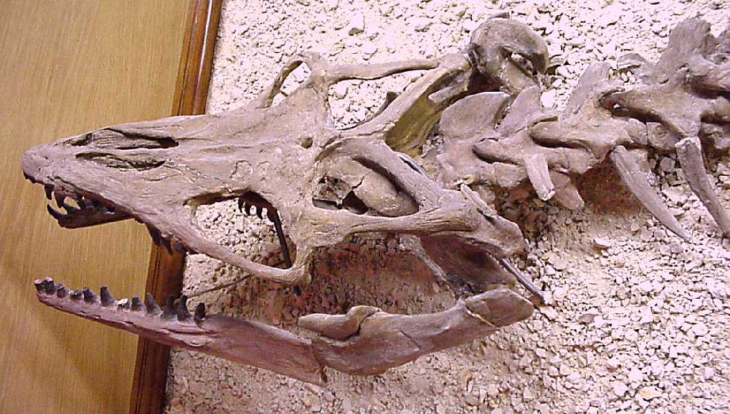
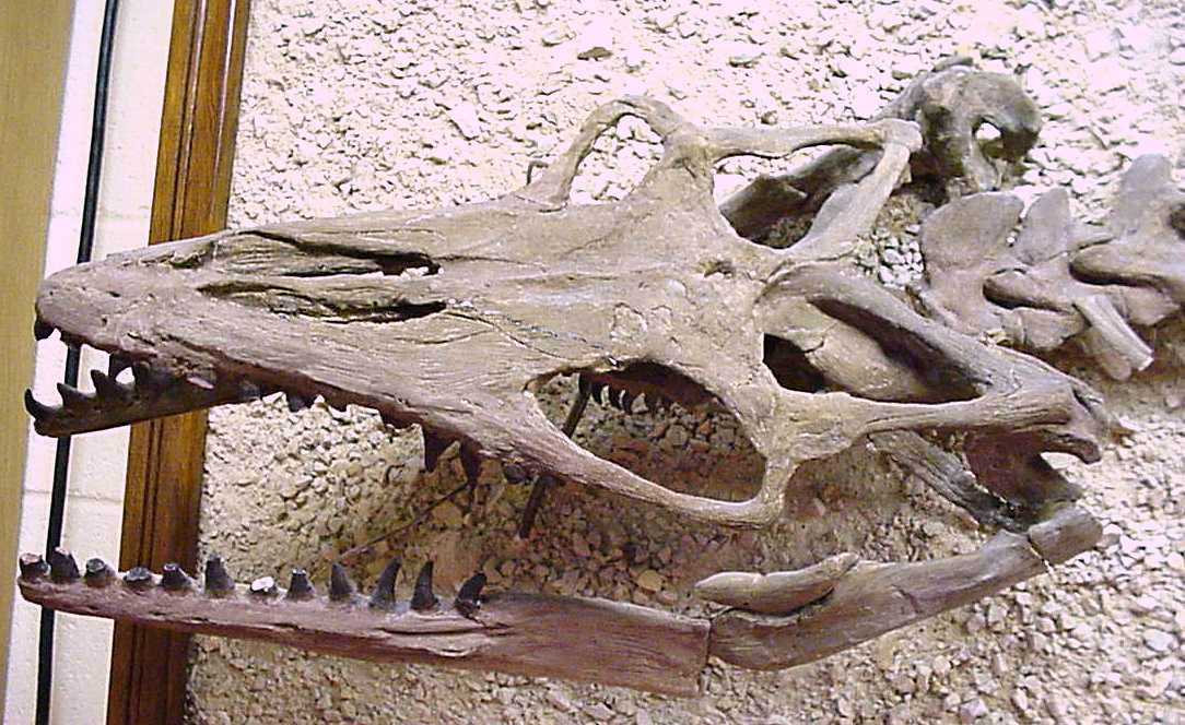
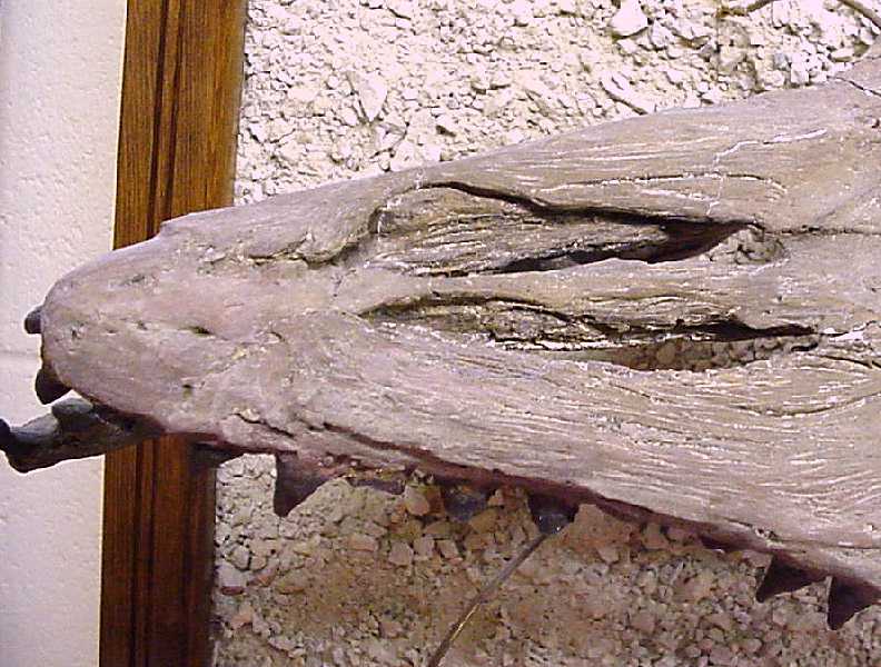
Platecarpus tympaniticus
skull,
dorsal view with lower jaws. This specimen is featured on the Platecarpus
webpage. NEW 2/2000 - For close-ups of the skull, go to Page11a. |
Sternberg Museum VP-17017 |
 |
NEW 2/2000 Platecarpus ictericus
skull, dorsal view |
Sternberg Museum VP-2077 |
 |
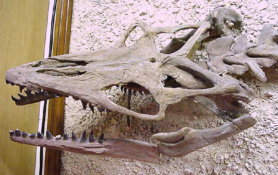
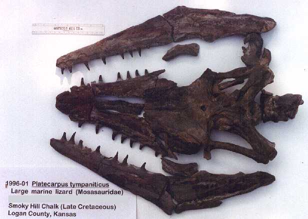
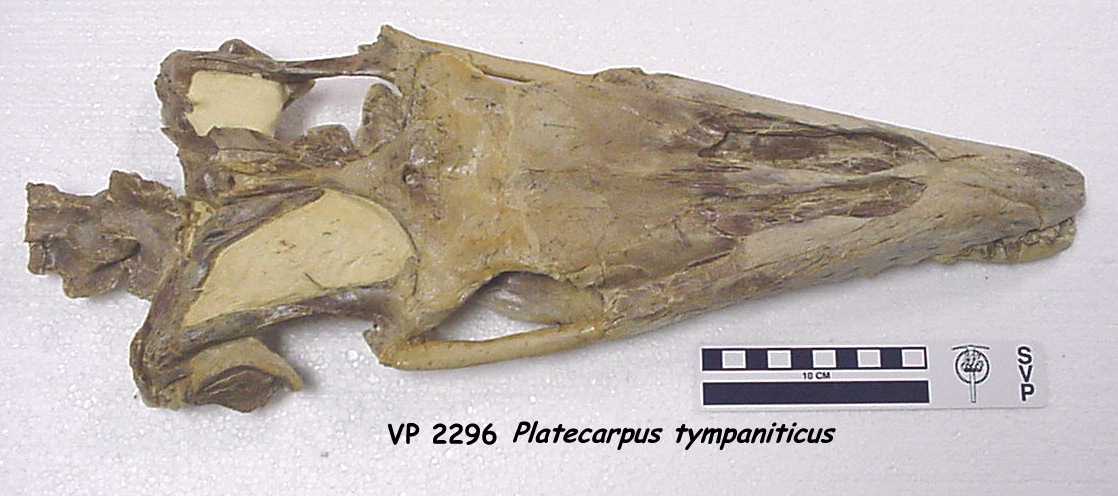
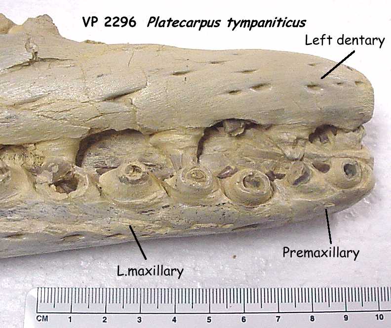
NEW 2/2000 Platecarpus planifrons skull,
dorsal view. For additional views of the skull, click here: Underside and detail
of jaws. |
Sternberg Museum VP-2296 |
 |
Platecarpus planifrons, skull, dorsal vertebra
and limb elements |
Sternberg
Museum VP-2116 |
 |
Platecarpus planifrons, dorsal view of the
skull. NEW 2/2000 |
Sternberg Museum VP-2116 |
 |
Platecarpus planifrons, New picture of the
dorsal side of the skull. |
Sternberg Museum VP-2116 |
 |
Platecarpus planifrons, dorsal
view of skull elements. This specimen is featured on the another
mosasaur discovery webpage. Click here for a picture of the assembled skull. |
Sternberg Museum VP-13010 |
 |
Platecarpus tympaniticus
, lateral view of premaxilla
and left maxilla. This specimen is featured on the another
mosasaur discovery webpage. Click here for a webpage describing the reconstruction of
this unique mosasaur skull. |
Sternberg Museum VP-13010 |
 |
Platecarpus tympaniticus, ventral view of muzzle unit
(premaxilla and maxillaries). This specimen is featured on the another
mosasaur discovery webpage. |
Sternberg Museum VP-13010 |
 |
Platecarpus tympaniticus, left and right
quadrates. This specimen is featured on the another mosasaur
discovery webpage. |
Sternberg Museum VP-13010 |
 |
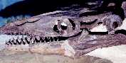
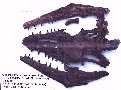
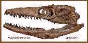
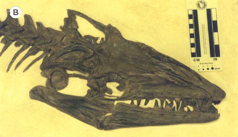
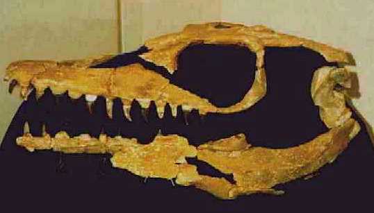
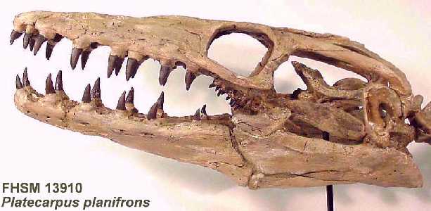
Platecarpus sp. skull, lateral view |
The Field Museum of
Natural History, Chicago, IL |
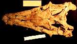 |
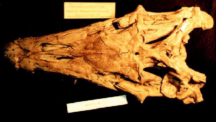
Platecarpus planifrons. skull, dorsal view of
complete skull, including lower jaws |
The University of Kansas, Museum of Natural History |
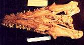 |
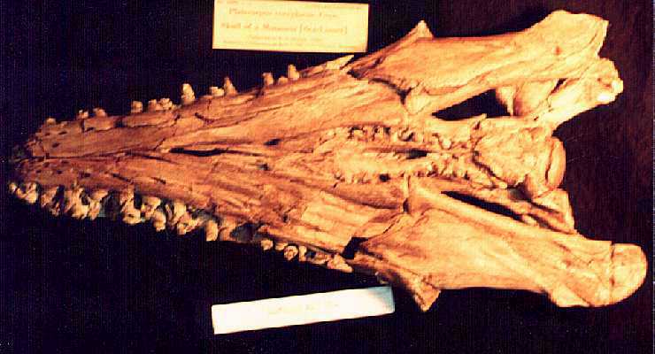
Platecarpus planifrons. skull, ventral view
of complete skull, including lower jaws |
The University of Kansas, Museum of Natural History |
 |
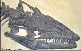
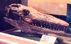
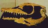
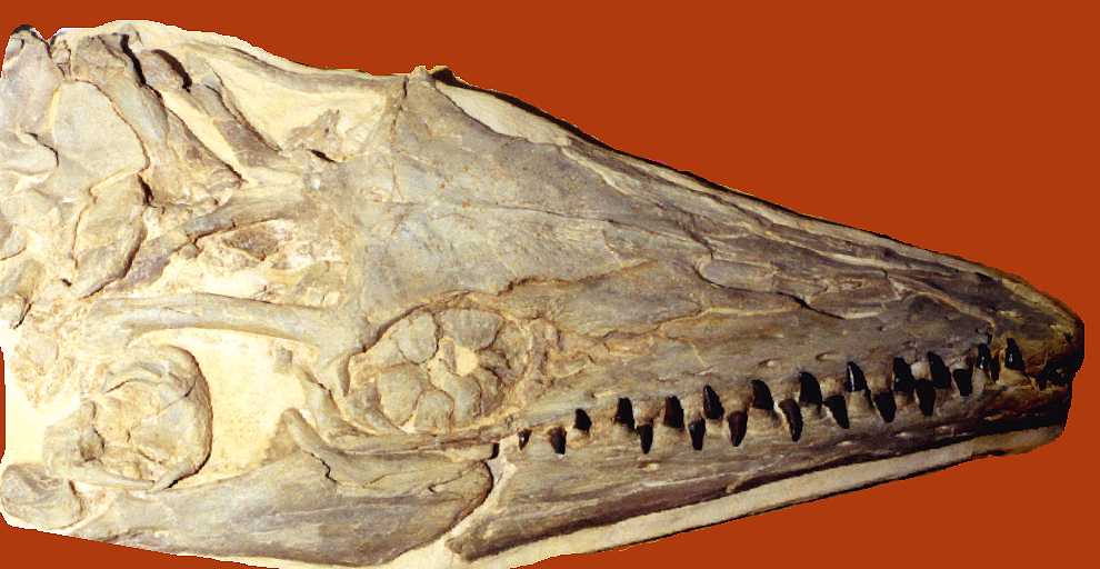
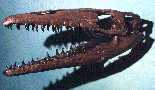
An intact and completely articulated Platecarpus
tympaniticus
skull found and prepared by Chuck Bonner. About 24 inches long. |
The Keystone
Gallery |
 |
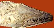
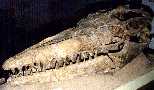
A fairly complete Platecarpus tympaniticus
skull from the Pierre Shale of South Dakota |
The Museum of Geology at the
South Dakota School of Mines and Technology |
 |
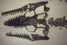
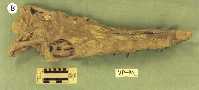
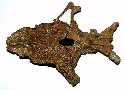
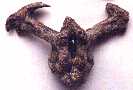
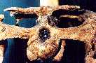
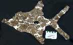
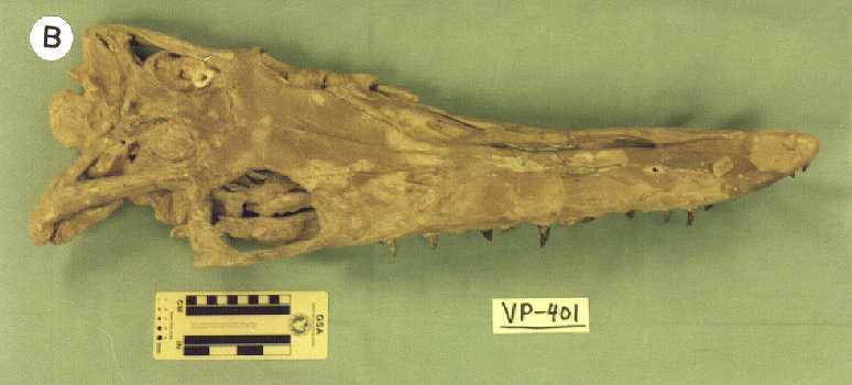
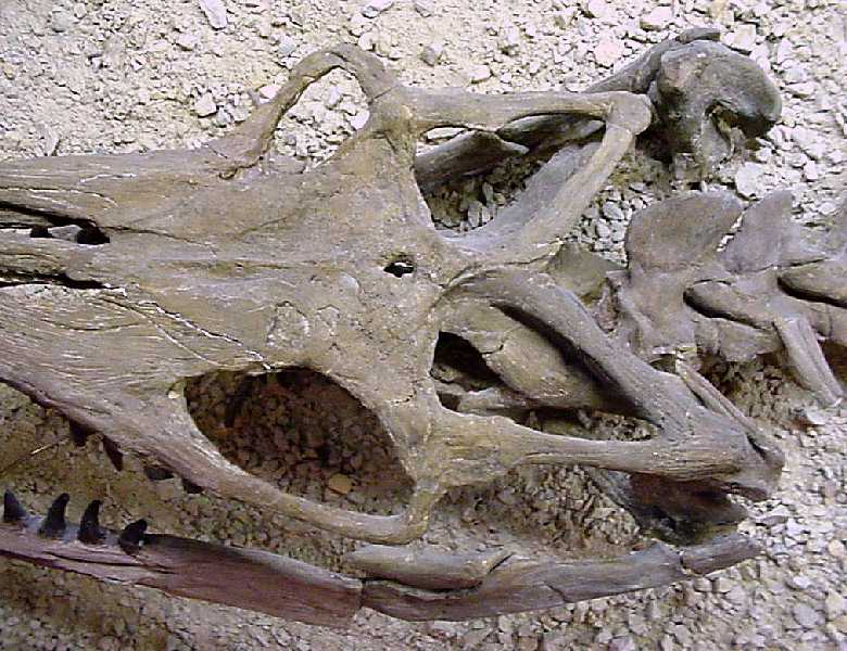
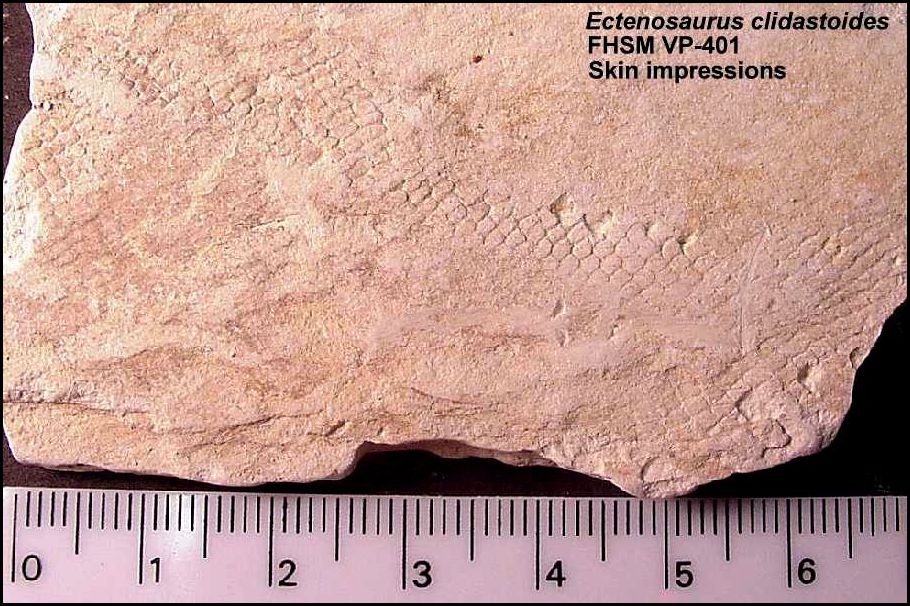
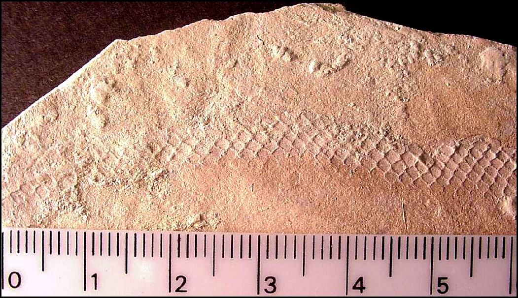
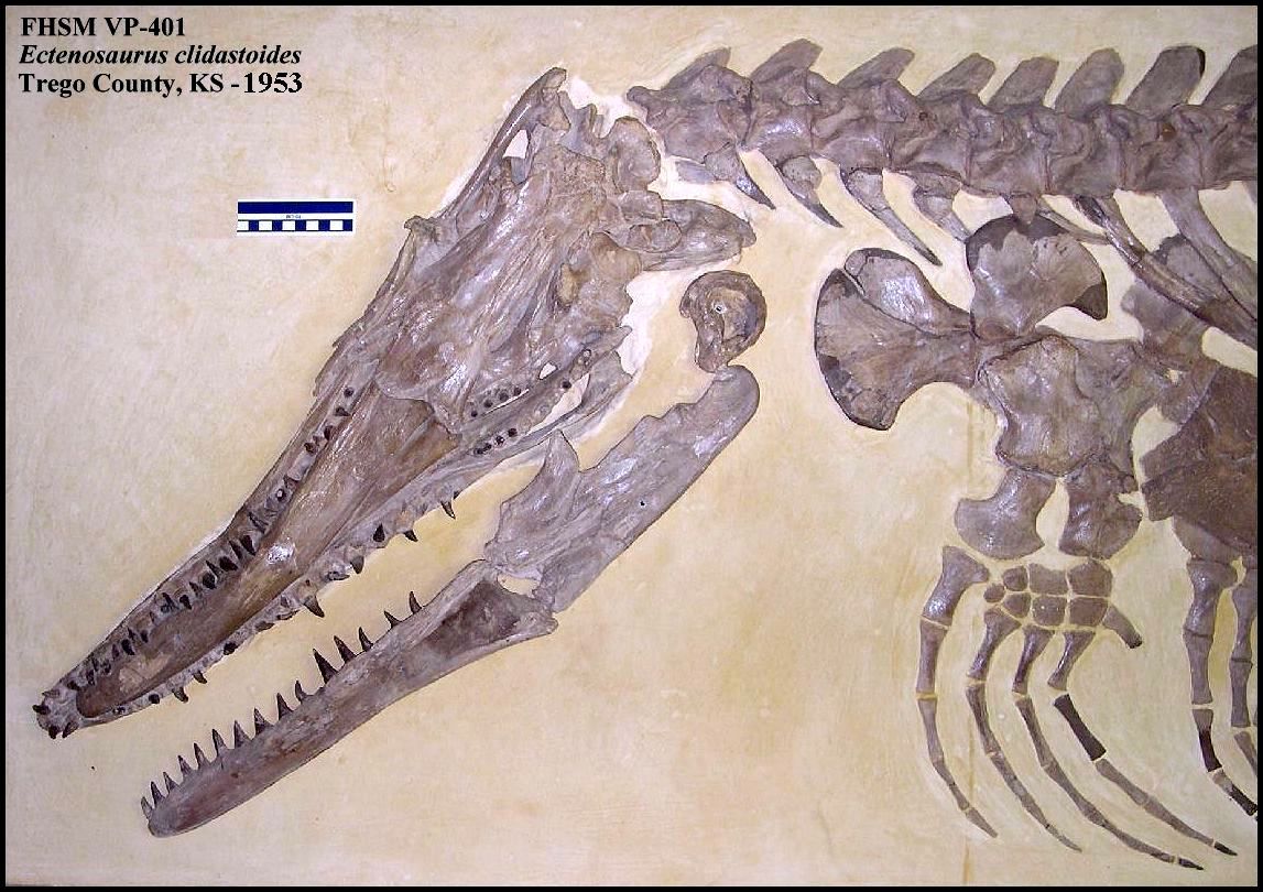
Ectenosaurus clidastoides (FHSM VP_401).
This specimen also preserved scale impressions: Scales 1
- Scales 2 - Scales
3 |
Sternberg Museum
VP-401 |
 |
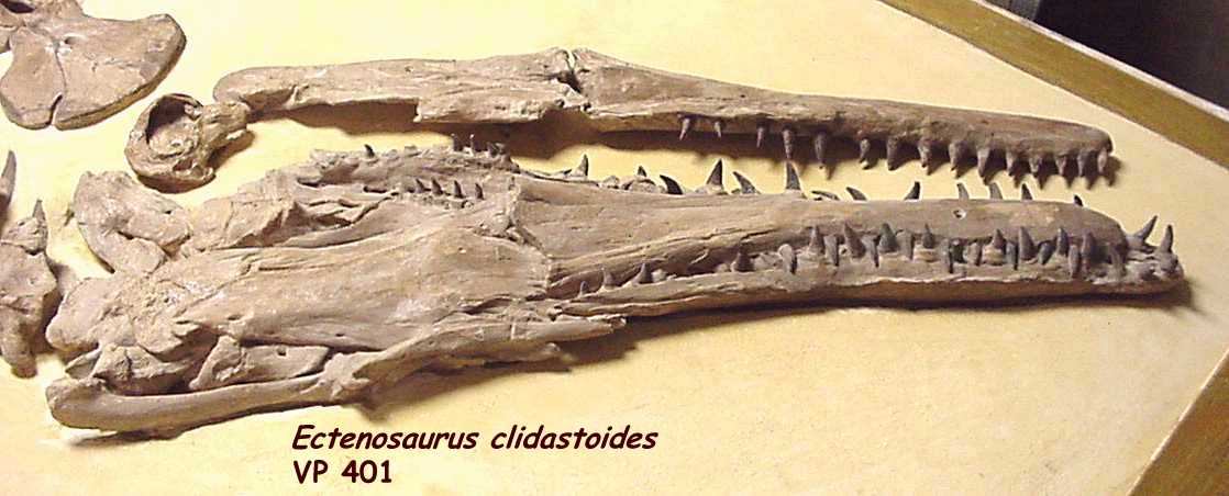
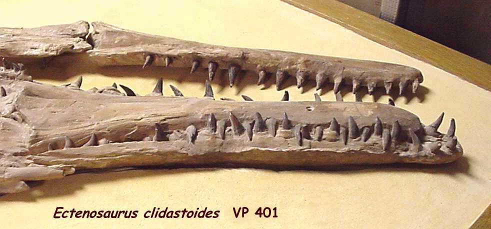
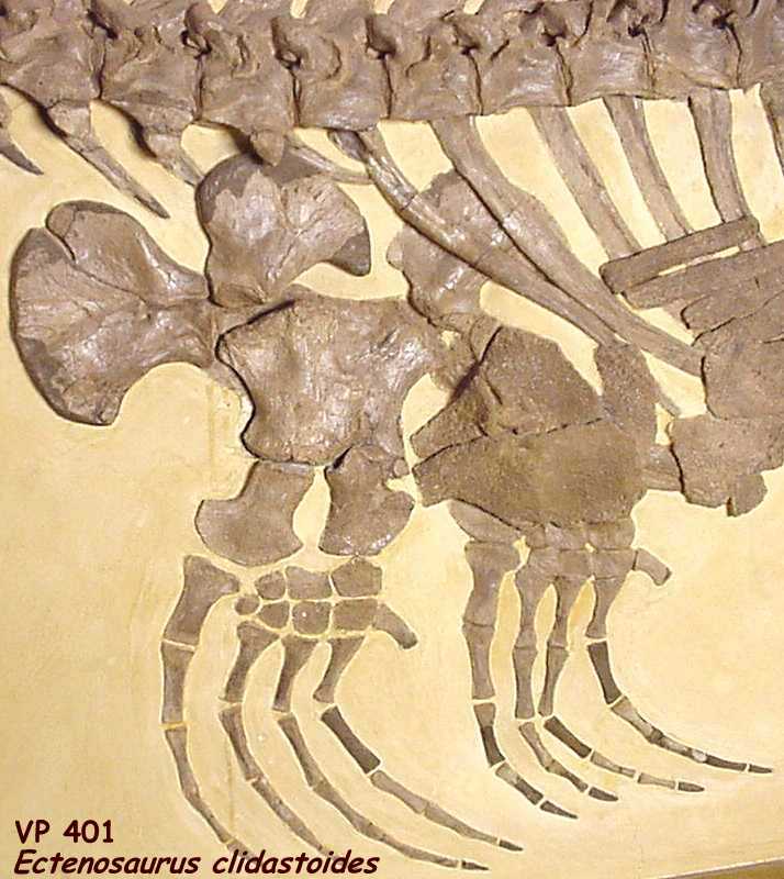
Ectenosaurus clidastoides, dorsal cranial view |
Sternberg Museum VP-401 |
 |
Ectenosaurus clidastoides, slightly different
lighting and angle. NEW 2/2000 - For additional pictures of this specimen, click
on the following: skull, jaws 1, jaws 2 and front limbs. |
Sternberg Museum VP-401 |
 |
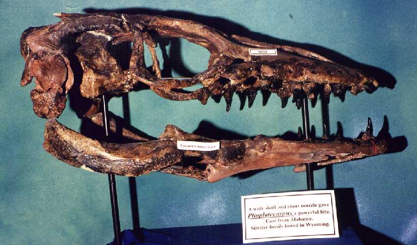
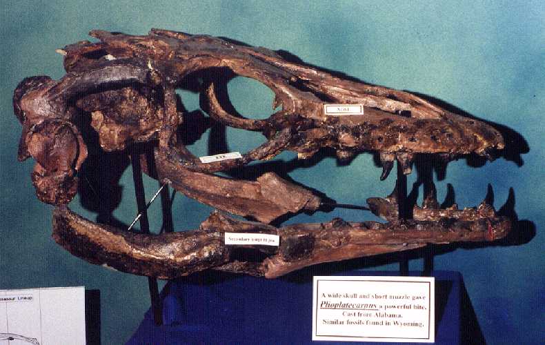
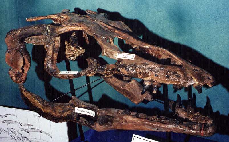
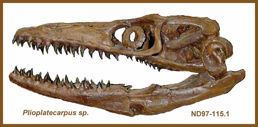
A lateral, left side view of a Plioplatecarpus sp.
skull from Alabama. This specimen is featured on the Plioplatecarpus
webpage. |
Okaloosa Walton Community College, Florida |
 |
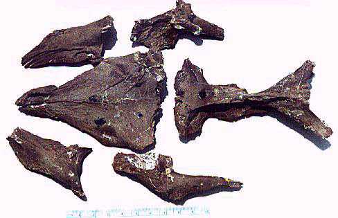
A dorsal view of the frontal, parietal and partial
post-orbitalfrontals from a recent mosasaur find in Lee County, Mississippi. The
remains have been tentatively identified as Plioplatecarpus primaevus, a medium
sized mosasaur. |
Private collection |
 |
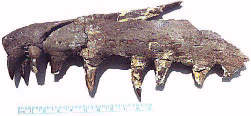
The muzzle of the above specimen, showing several well
preserved teeth. This specimen comes from the Bluffport Marl member of the Demopolis Chalk, a formation that was deposited in the
Mississippian Embayment. |
Private collection |
 |
A dorsal view of the parietal and frontal of the only Plioplatecarpus found to date in Kansas. This
picture shows the large parietal foremen which is a characteristic of Plioplatecarpus.
Check out the Kansas Plioplatecarpus site for
additional pictures |
Private collection |
 |
A ventral view of the parietal (left) and frontal
(right) of the same specimen. Although these remains were partially prepared, the invasion
of selenite crystals into the bone has already destroyed much of the detail of the surface |
Private collection |
 |
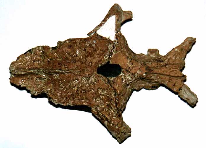
The braincase of this Plioplatecarpus
specimen, from above (the basio-occipital and basio- splenoid bones that form the base of
the braincase (center) are flanked by the left and right pro-otics. |
Private collection |
 |
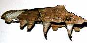
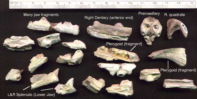
Pieces of the lower jaws of the Plioplatecarpus
sp. specimen from the upper portion of the Sharon Springs member of the Pierre Shale,
Logan County, KS. The teeth were still in surprising good shape for a 'shale
fossil'. |
Private collection |
 |
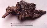
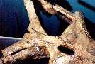
An articulated series of four cervical vertebrae. |
Private collection |
 |
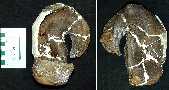
From left to right, an exterior view of the left
quadrate, and an interior view of the right quadrate of Plioplatecarpus.
Note the stapedial pit on the inside of the right quadrate. This structure is useful
in determining the species of the mosasaur |
Private collection |
 |
Two pathological dorsal vertebrae (fused) showing
evidence of a healed spinal injury or disease that must have been painful, and may have
seriously hindered the swimming ability of this mosasaur. |
Private collection |
 |
A nicely preserved dorsal vertebrae. This
specimen is of late Campanian age and is comparable with material from the lower Pierre
Shale of Kansas and S. Dakota. |
Private collection |
 |
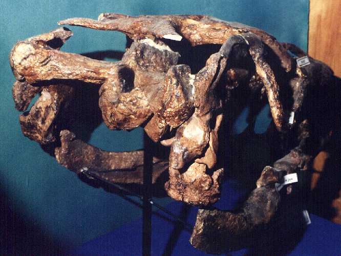
Head on view of the cast of a large Plioplatecarpus
skull (UNO 8611-2). The original specimen was prepared and described by David
Burnham in his Master's thesis at the University of New Orleans, 1991. |
Exhibited in the Tate Geological Museum, Casper,
WY |
 |
Right side view of the same skull, showing the
unusually large eyes and shorter snout that are typical of Plioplatecarpus.
The original specimen was found in the Demopolis
Chalk of western Alabama. |
Exhibited in the Tate Geological Museum, Casper, WY |
 |
Another right side view of the same skull
Several drawings of this nearly complete skull by David Burnham are shown on the Plioplatecarpus webpage. |
Exhibited in the Tate Geological Museum, Casper, WY |
 |
Plioplatecarpus was one of several mosasaurs
that were still around at the end the Cretaceous and occur somewhat later in the geologic
record than the mosasaurs from the Smoky Hill Chalk. |
Exhibited in the Tate Geological Museum, Casper,
WY |
 |
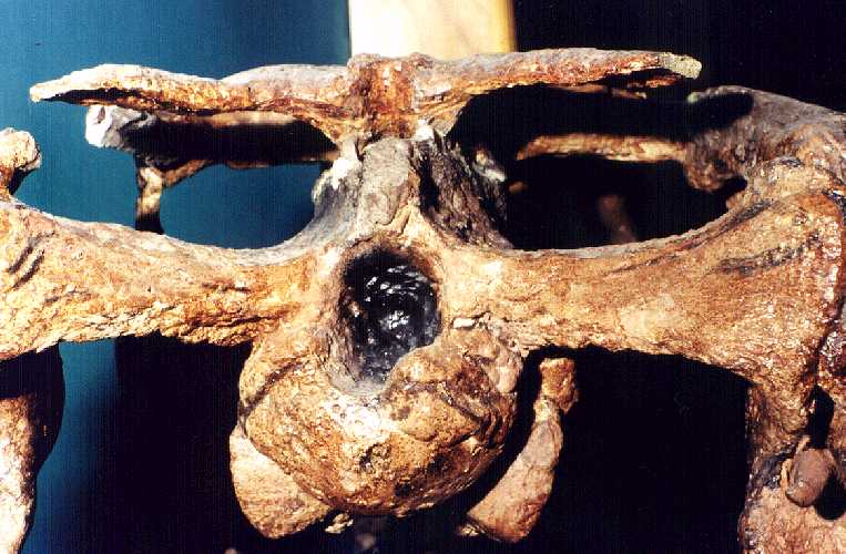
A view of the back of the skull, showing the occipital
condyle where the first vertebra is attached, and the opening for the spinal cord into the
braincase. |
Exhibited in the Tate Geological Museum, Casper, WY |
 |
Another rear view of the skull, showing the occipital
condyle, the size of the braincase, and the open framework of the skull. In life,
these openings were filled with the strong muscles that closed the jaw. |
Exhibited in the Tate Geological Museum, Casper, WY |
 |
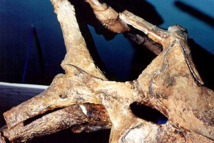
A view of the top of the skull (and the suture between
the parietal and frontal bones) showing the large parietal foremen that is typical of
plioplatecarpines. The function of this opening in mosasaurs is not known. |
Exhibited in the Tate Geological Museum, Casper, WY |
 |
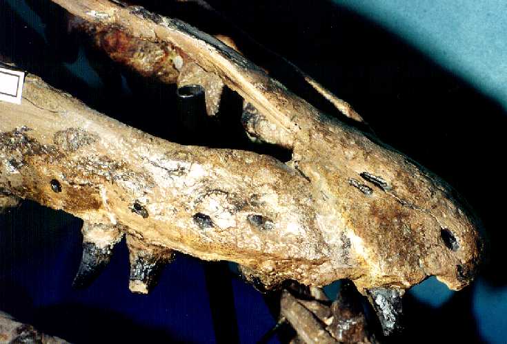
The snout, showing the suture between the premaxilla
bone (top) and the right maxilla. The nasal openings are relatively larger in Plioplatecarpus
compared to other mosasaurs. |
Exhibited in the Tate Geological Museum, Casper, WY |
 |
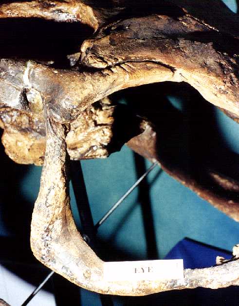
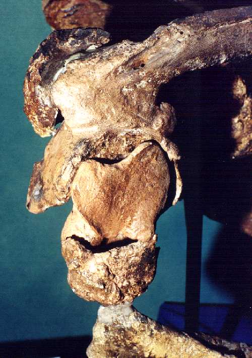
The orbit of the right eye, formed by the jugal bone
below and behind, the post orbital frontal above, the frontal, and the prefrontal.
The scratches over the eye appear to be bite marks, possibly caused by scavenging. |
Exhibited in the Tate Geological Museum, Casper, WY |
 |
A view of the right quadrate. Some damage to the
joint at the back of the right lower jaw is apparent. |
Exhibited in the Tate Geological Museum, Casper,
WY |
 |
Possibly a new species of Plioplatecarpus
from North Dakota, this animal was larger than previous known specimens. For more
information see the North Dakota Plioplatecarpus page. |
Soon to be on exhibit in the North Dakota Heritage Center
(ND97-115.1) |
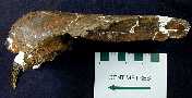 |
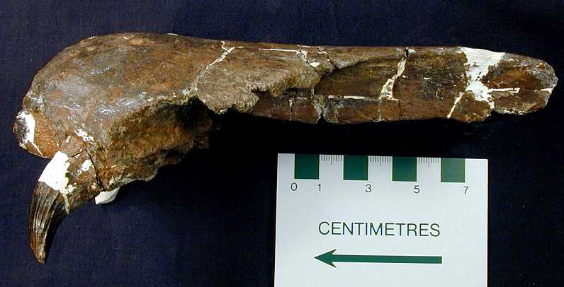
A left side view of the premaxilla or muzzle of the
mosasaur. Scale is in centimeters (cm). Pictures courtesy of John Campbell, North Dakota Geological Survey. |
Soon to be on exhibit in the North Dakota Heritage
Center (ND97-115.1) |
 |
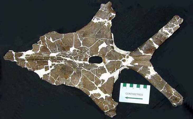
A dorsal view of the frontal and parietal bones that
comprise the top of the skull. |
Soon to be on exhibit in the North Dakota Heritage Center
(ND97-115.1) |
 |
A lateral and medial view of the left quadrate of the
mosasaur. Pictures courtesy of John Campbell, North
Dakota Geological Survey. |
Soon to be on exhibit in the North Dakota Heritage
Center (ND97-115.1) |
 |
Bite marks on the right jugal (lower and rear part of
the orbit of the eye) which indicate scavenging of the carcass by sharks. |
Soon to be on exhibit in the North Dakota Heritage Center (ND97-115.1) |
 |
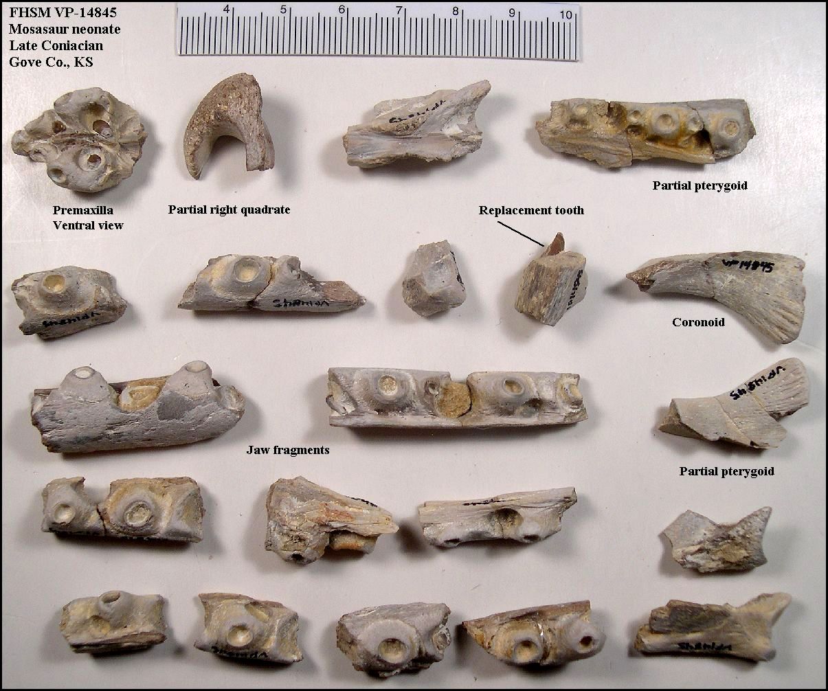
The fragmentary skull of a very small mosasaur
(possibly a Platecarpus unborn fetus or recently born neonate) from the lower
Smoky Hill Chalk of Kansas. |
Sternberg Museum VP-14845 |
 |
An updated photograph of the above specimen (after
donation). |
Sternberg Museum VP-14845 |


